{"title":"非典型胃病变提示潜在胰腺癌直接侵袭。","authors":"Byeong Joo Jo","doi":"10.7704/kjhugr.2025.0032","DOIUrl":null,"url":null,"abstract":"<p><p>Atypical gastric lesions present diagnostic challenges, particularly when they mimic benign conditions. We report the case of a 76-year-old male who underwent esophagogastroduodenoscopy (EGD) during routine screening. The EGD revealed a 1-cm ulcerative lesion in the lesser curvature of the stomach. The initial endoscopic report simply described the lesion as a \"gastric ulcer,\" and the initial biopsy indicated a tubulovillous adenoma with low-grade dysplasia. However, follow-up endoscopy demonstrated an irregular, non-healing ulcer without a clean base, raising a strong possibility of deeper infiltration. A follow-up biopsy showed atypical findings, suggestive of malignancy, and prompting further evaluation. Abdominal computed tomography (CT) revealed a mass in the pancreatic body and tail that directly invaded the gastric wall, along with extensive peritoneal metastasis. Positron emission tomography-CT confirmed hypermetabolic activity in the pancreatic lesion. The final diagnosis was a pancreatic adenocarcinoma with gastric invasion. This case highlights the importance of correlating endoscopic findings with imaging and pathology findings, especially when endoscopic morphology appears atypical and inconsistent with biopsy results. Repeat evaluations and cross-sectional imaging may prevent unnecessary procedures and allow timely identification of extrinsic malignancies that present as primary gastric lesions.</p>","PeriodicalId":520887,"journal":{"name":"The Korean journal of helicobacter and upper gastrointestinal research","volume":"25 3","pages":"288-291"},"PeriodicalIF":0.0000,"publicationDate":"2025-09-01","publicationTypes":"Journal Article","fieldsOfStudy":null,"isOpenAccess":false,"openAccessPdf":"https://www.ncbi.nlm.nih.gov/pmc/articles/PMC12425658/pdf/","citationCount":"0","resultStr":"{\"title\":\"Atypical Gastric Lesion Revealing Underlying Pancreatic Cancer With Direct Invasion.\",\"authors\":\"Byeong Joo Jo\",\"doi\":\"10.7704/kjhugr.2025.0032\",\"DOIUrl\":null,\"url\":null,\"abstract\":\"<p><p>Atypical gastric lesions present diagnostic challenges, particularly when they mimic benign conditions. We report the case of a 76-year-old male who underwent esophagogastroduodenoscopy (EGD) during routine screening. The EGD revealed a 1-cm ulcerative lesion in the lesser curvature of the stomach. The initial endoscopic report simply described the lesion as a \\\"gastric ulcer,\\\" and the initial biopsy indicated a tubulovillous adenoma with low-grade dysplasia. However, follow-up endoscopy demonstrated an irregular, non-healing ulcer without a clean base, raising a strong possibility of deeper infiltration. A follow-up biopsy showed atypical findings, suggestive of malignancy, and prompting further evaluation. Abdominal computed tomography (CT) revealed a mass in the pancreatic body and tail that directly invaded the gastric wall, along with extensive peritoneal metastasis. Positron emission tomography-CT confirmed hypermetabolic activity in the pancreatic lesion. The final diagnosis was a pancreatic adenocarcinoma with gastric invasion. This case highlights the importance of correlating endoscopic findings with imaging and pathology findings, especially when endoscopic morphology appears atypical and inconsistent with biopsy results. Repeat evaluations and cross-sectional imaging may prevent unnecessary procedures and allow timely identification of extrinsic malignancies that present as primary gastric lesions.</p>\",\"PeriodicalId\":520887,\"journal\":{\"name\":\"The Korean journal of helicobacter and upper gastrointestinal research\",\"volume\":\"25 3\",\"pages\":\"288-291\"},\"PeriodicalIF\":0.0000,\"publicationDate\":\"2025-09-01\",\"publicationTypes\":\"Journal Article\",\"fieldsOfStudy\":null,\"isOpenAccess\":false,\"openAccessPdf\":\"https://www.ncbi.nlm.nih.gov/pmc/articles/PMC12425658/pdf/\",\"citationCount\":\"0\",\"resultStr\":null,\"platform\":\"Semanticscholar\",\"paperid\":null,\"PeriodicalName\":\"The Korean journal of helicobacter and upper gastrointestinal research\",\"FirstCategoryId\":\"1085\",\"ListUrlMain\":\"https://doi.org/10.7704/kjhugr.2025.0032\",\"RegionNum\":0,\"RegionCategory\":null,\"ArticlePicture\":[],\"TitleCN\":null,\"AbstractTextCN\":null,\"PMCID\":null,\"EPubDate\":\"\",\"PubModel\":\"\",\"JCR\":\"\",\"JCRName\":\"\",\"Score\":null,\"Total\":0}","platform":"Semanticscholar","paperid":null,"PeriodicalName":"The Korean journal of helicobacter and upper gastrointestinal research","FirstCategoryId":"1085","ListUrlMain":"https://doi.org/10.7704/kjhugr.2025.0032","RegionNum":0,"RegionCategory":null,"ArticlePicture":[],"TitleCN":null,"AbstractTextCN":null,"PMCID":null,"EPubDate":"","PubModel":"","JCR":"","JCRName":"","Score":null,"Total":0}
Atypical Gastric Lesion Revealing Underlying Pancreatic Cancer With Direct Invasion.
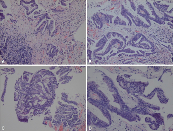
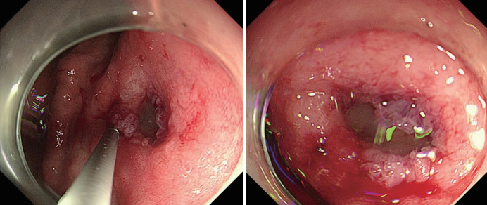
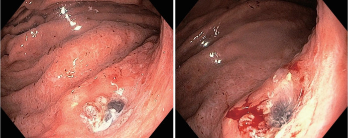
Atypical gastric lesions present diagnostic challenges, particularly when they mimic benign conditions. We report the case of a 76-year-old male who underwent esophagogastroduodenoscopy (EGD) during routine screening. The EGD revealed a 1-cm ulcerative lesion in the lesser curvature of the stomach. The initial endoscopic report simply described the lesion as a "gastric ulcer," and the initial biopsy indicated a tubulovillous adenoma with low-grade dysplasia. However, follow-up endoscopy demonstrated an irregular, non-healing ulcer without a clean base, raising a strong possibility of deeper infiltration. A follow-up biopsy showed atypical findings, suggestive of malignancy, and prompting further evaluation. Abdominal computed tomography (CT) revealed a mass in the pancreatic body and tail that directly invaded the gastric wall, along with extensive peritoneal metastasis. Positron emission tomography-CT confirmed hypermetabolic activity in the pancreatic lesion. The final diagnosis was a pancreatic adenocarcinoma with gastric invasion. This case highlights the importance of correlating endoscopic findings with imaging and pathology findings, especially when endoscopic morphology appears atypical and inconsistent with biopsy results. Repeat evaluations and cross-sectional imaging may prevent unnecessary procedures and allow timely identification of extrinsic malignancies that present as primary gastric lesions.

 求助内容:
求助内容: 应助结果提醒方式:


