{"title":"计算机断层扫描、超声和阴道镜检查在绝育雌性拉布拉多犬双叶状阴道<s:1>勒氏囊肿中的应用。","authors":"Ying-Ying Lo, Emmanuel Topie, Stéphanie Moreau, Dimitri Leperlier, Anne-Sophie Bedu","doi":"10.1155/crve/5706632","DOIUrl":null,"url":null,"abstract":"<p><p>An 11-year-old neutered female Labrador was presented with dyschezia and dysuria associated with a large perineal mass that had been present for 2 years. Computed tomography (CT) revealed a large bilobed, cavitated mass localized ventrally to the rectum and dorsally to the urethra at the level of the vagina, with hypoattenuating contents and a contrast-enhancing peripheral wall. Surgical excision was performed and confirmed the cystic nature of the mass. Histopathological findings were compatible with a benign vaginal cyst, most likely originating from the Müllerian or paramesonephric ducts. The CT provided relevant information for surgical planning and enabled accurate assessment of the mass's location, extent, and relationship with adjacent structures.</p>","PeriodicalId":37339,"journal":{"name":"Case Reports in Veterinary Medicine","volume":"2025 ","pages":"5706632"},"PeriodicalIF":0.0000,"publicationDate":"2025-09-02","publicationTypes":"Journal Article","fieldsOfStudy":null,"isOpenAccess":false,"openAccessPdf":"https://www.ncbi.nlm.nih.gov/pmc/articles/PMC12419931/pdf/","citationCount":"0","resultStr":"{\"title\":\"Use of Computed Tomography, Ultrasound, and Vaginoscopy in the Evaluation of a Bilobed Vaginal Müllerian Cyst in a Neutered Female Labrador Retriever.\",\"authors\":\"Ying-Ying Lo, Emmanuel Topie, Stéphanie Moreau, Dimitri Leperlier, Anne-Sophie Bedu\",\"doi\":\"10.1155/crve/5706632\",\"DOIUrl\":null,\"url\":null,\"abstract\":\"<p><p>An 11-year-old neutered female Labrador was presented with dyschezia and dysuria associated with a large perineal mass that had been present for 2 years. Computed tomography (CT) revealed a large bilobed, cavitated mass localized ventrally to the rectum and dorsally to the urethra at the level of the vagina, with hypoattenuating contents and a contrast-enhancing peripheral wall. Surgical excision was performed and confirmed the cystic nature of the mass. Histopathological findings were compatible with a benign vaginal cyst, most likely originating from the Müllerian or paramesonephric ducts. The CT provided relevant information for surgical planning and enabled accurate assessment of the mass's location, extent, and relationship with adjacent structures.</p>\",\"PeriodicalId\":37339,\"journal\":{\"name\":\"Case Reports in Veterinary Medicine\",\"volume\":\"2025 \",\"pages\":\"5706632\"},\"PeriodicalIF\":0.0000,\"publicationDate\":\"2025-09-02\",\"publicationTypes\":\"Journal Article\",\"fieldsOfStudy\":null,\"isOpenAccess\":false,\"openAccessPdf\":\"https://www.ncbi.nlm.nih.gov/pmc/articles/PMC12419931/pdf/\",\"citationCount\":\"0\",\"resultStr\":null,\"platform\":\"Semanticscholar\",\"paperid\":null,\"PeriodicalName\":\"Case Reports in Veterinary Medicine\",\"FirstCategoryId\":\"1085\",\"ListUrlMain\":\"https://doi.org/10.1155/crve/5706632\",\"RegionNum\":0,\"RegionCategory\":null,\"ArticlePicture\":[],\"TitleCN\":null,\"AbstractTextCN\":null,\"PMCID\":null,\"EPubDate\":\"2025/1/1 0:00:00\",\"PubModel\":\"eCollection\",\"JCR\":\"Q3\",\"JCRName\":\"Veterinary\",\"Score\":null,\"Total\":0}","platform":"Semanticscholar","paperid":null,"PeriodicalName":"Case Reports in Veterinary Medicine","FirstCategoryId":"1085","ListUrlMain":"https://doi.org/10.1155/crve/5706632","RegionNum":0,"RegionCategory":null,"ArticlePicture":[],"TitleCN":null,"AbstractTextCN":null,"PMCID":null,"EPubDate":"2025/1/1 0:00:00","PubModel":"eCollection","JCR":"Q3","JCRName":"Veterinary","Score":null,"Total":0}
Use of Computed Tomography, Ultrasound, and Vaginoscopy in the Evaluation of a Bilobed Vaginal Müllerian Cyst in a Neutered Female Labrador Retriever.
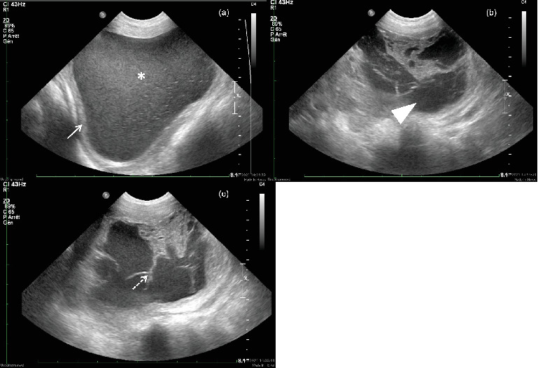
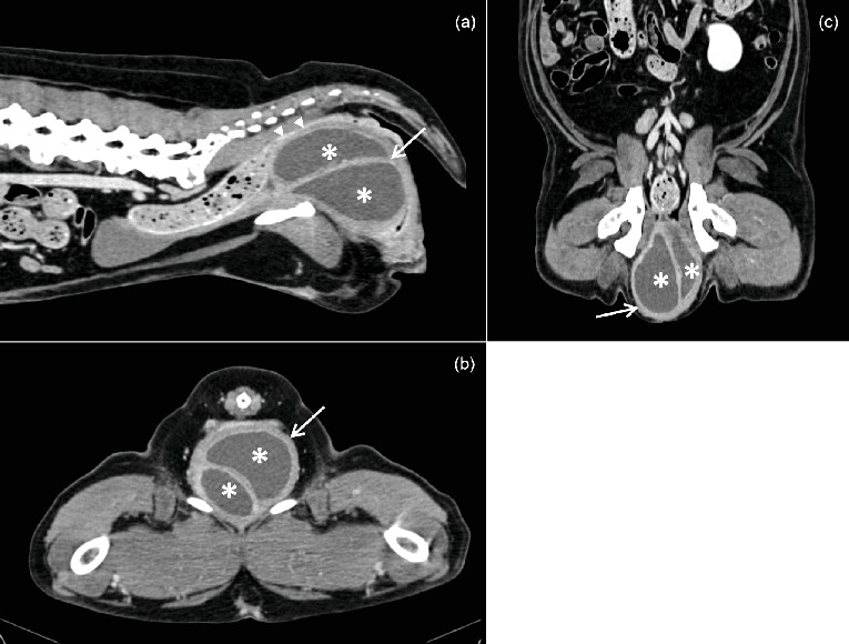
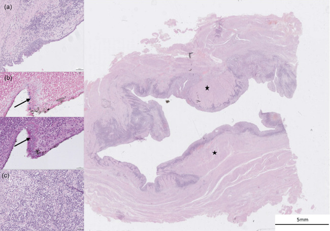
An 11-year-old neutered female Labrador was presented with dyschezia and dysuria associated with a large perineal mass that had been present for 2 years. Computed tomography (CT) revealed a large bilobed, cavitated mass localized ventrally to the rectum and dorsally to the urethra at the level of the vagina, with hypoattenuating contents and a contrast-enhancing peripheral wall. Surgical excision was performed and confirmed the cystic nature of the mass. Histopathological findings were compatible with a benign vaginal cyst, most likely originating from the Müllerian or paramesonephric ducts. The CT provided relevant information for surgical planning and enabled accurate assessment of the mass's location, extent, and relationship with adjacent structures.
期刊介绍:
Case Reports in Veterinary Medicine is a peer-reviewed, Open Access journal that publishes case reports and case series in all areas of veterinary medicine.

 求助内容:
求助内容: 应助结果提醒方式:


