Sara Boccalini, Clara Fourrier, Salim Si-Mohamed, Eric Bonnefoy-Cudraz, Thomas Bochaton, Loic Boussel, Anna Vlachomitrou, Rafael Wiemker, Philippe Douek
{"title":"心肌炎病变的频谱CT表现及其与MRI水肿的相关性。","authors":"Sara Boccalini, Clara Fourrier, Salim Si-Mohamed, Eric Bonnefoy-Cudraz, Thomas Bochaton, Loic Boussel, Anna Vlachomitrou, Rafael Wiemker, Philippe Douek","doi":"10.1186/s41747-025-00613-x","DOIUrl":null,"url":null,"abstract":"<p><strong>Background: </strong>Spectral computed tomography (CT) late-enhancement (LE) acquisitions can help detect myocarditis. An arterial acquisition is often performed for coronary artery analysis. However, little is known about the appearance of myocarditis on the arterial phase. We investigated the appearance of myocarditis on arterial acquisitions of cardiac spectral CT, and its relationship to LE and edema.</p><p><strong>Materials and methods: </strong>Forty-seven cardiac spectral CTs performed in patients with magnetic resonance imaging (MRI)-confirmed myocarditis were retrospectively assessed. Three myocardial attenuation/enhancement patterns were visually identified and segmented on both arterial and LE acquisitions: hypodense-arterial + normal-LE (HypoArt-NorLE); normal-arterial + hyperdense-LE (NorArt-HyperLE); and hypodense-arterial + hyperdense-late (HypoArt-HyperLE). Characteristics of conventional and spectral images were calculated for all patterns and for remote myocardium. Values of HypoArt-HyperLE lesions were compared in the groups with and without edema on MRI, as assessed with T2 mapping (available for 25 patients).</p><p><strong>Results: </strong>We found 173 lesions, 46 (26%) HypoArt-NorLE, 54 (31%) NorArt-HyperLE, and 73 (42%) HypoArt-HyperLE. On the arterial phase, HypoArt-HyperLE were more hypodense (p < 0.001) and had less iodine (0.23 mg/mL less; p < 0.001) than RM. On LE, both HypoArt-HyperLE and NorArt-HyperLE were more hyperdense and contained more iodine than the remote myocardium (all p < 0.001). HypoArt-HyperLE lesions were more hypodense and contained less iodine on the arterial phase in patients with edema on MRI as compared to those without (all p < 0.001).</p><p><strong>Conclusion: </strong>Most myocarditis lesions detectable with spectral CT are visible on both arterial and LE acquisitions. These lesions appeared to be more pronounced on the arterial phase in patients with edema on MRI.</p><p><strong>Relevance statement: </strong>Spectral CT arterial acquisition performed for the differential diagnosis of acute myocardial pathologies in many cases can depict myocarditis lesions as epicardial hypodense areas, most likely related to the presence of edema.</p><p><strong>Key points: </strong>Data from spectral CT shows that most myocarditis lesions appear as hypodense on the arterial phase, matching the epicardial LE zones. A minority of myocarditis lesions appear as epicardial LE areas without anomalies of attenuation on the arterial phase. Hypodense myocardial areas are correlated to the presence of edema on MRI, suggesting they are due to the same phenomenon.</p>","PeriodicalId":36926,"journal":{"name":"European Radiology Experimental","volume":"9 1","pages":"90"},"PeriodicalIF":3.6000,"publicationDate":"2025-09-11","publicationTypes":"Journal Article","fieldsOfStudy":null,"isOpenAccess":false,"openAccessPdf":"https://www.ncbi.nlm.nih.gov/pmc/articles/PMC12425864/pdf/","citationCount":"0","resultStr":"{\"title\":\"Appearance of myocarditis lesions on spectral CT arterial acquisitions and correlation with edema on MRI.\",\"authors\":\"Sara Boccalini, Clara Fourrier, Salim Si-Mohamed, Eric Bonnefoy-Cudraz, Thomas Bochaton, Loic Boussel, Anna Vlachomitrou, Rafael Wiemker, Philippe Douek\",\"doi\":\"10.1186/s41747-025-00613-x\",\"DOIUrl\":null,\"url\":null,\"abstract\":\"<p><strong>Background: </strong>Spectral computed tomography (CT) late-enhancement (LE) acquisitions can help detect myocarditis. An arterial acquisition is often performed for coronary artery analysis. However, little is known about the appearance of myocarditis on the arterial phase. We investigated the appearance of myocarditis on arterial acquisitions of cardiac spectral CT, and its relationship to LE and edema.</p><p><strong>Materials and methods: </strong>Forty-seven cardiac spectral CTs performed in patients with magnetic resonance imaging (MRI)-confirmed myocarditis were retrospectively assessed. Three myocardial attenuation/enhancement patterns were visually identified and segmented on both arterial and LE acquisitions: hypodense-arterial + normal-LE (HypoArt-NorLE); normal-arterial + hyperdense-LE (NorArt-HyperLE); and hypodense-arterial + hyperdense-late (HypoArt-HyperLE). Characteristics of conventional and spectral images were calculated for all patterns and for remote myocardium. Values of HypoArt-HyperLE lesions were compared in the groups with and without edema on MRI, as assessed with T2 mapping (available for 25 patients).</p><p><strong>Results: </strong>We found 173 lesions, 46 (26%) HypoArt-NorLE, 54 (31%) NorArt-HyperLE, and 73 (42%) HypoArt-HyperLE. On the arterial phase, HypoArt-HyperLE were more hypodense (p < 0.001) and had less iodine (0.23 mg/mL less; p < 0.001) than RM. On LE, both HypoArt-HyperLE and NorArt-HyperLE were more hyperdense and contained more iodine than the remote myocardium (all p < 0.001). HypoArt-HyperLE lesions were more hypodense and contained less iodine on the arterial phase in patients with edema on MRI as compared to those without (all p < 0.001).</p><p><strong>Conclusion: </strong>Most myocarditis lesions detectable with spectral CT are visible on both arterial and LE acquisitions. These lesions appeared to be more pronounced on the arterial phase in patients with edema on MRI.</p><p><strong>Relevance statement: </strong>Spectral CT arterial acquisition performed for the differential diagnosis of acute myocardial pathologies in many cases can depict myocarditis lesions as epicardial hypodense areas, most likely related to the presence of edema.</p><p><strong>Key points: </strong>Data from spectral CT shows that most myocarditis lesions appear as hypodense on the arterial phase, matching the epicardial LE zones. A minority of myocarditis lesions appear as epicardial LE areas without anomalies of attenuation on the arterial phase. Hypodense myocardial areas are correlated to the presence of edema on MRI, suggesting they are due to the same phenomenon.</p>\",\"PeriodicalId\":36926,\"journal\":{\"name\":\"European Radiology Experimental\",\"volume\":\"9 1\",\"pages\":\"90\"},\"PeriodicalIF\":3.6000,\"publicationDate\":\"2025-09-11\",\"publicationTypes\":\"Journal Article\",\"fieldsOfStudy\":null,\"isOpenAccess\":false,\"openAccessPdf\":\"https://www.ncbi.nlm.nih.gov/pmc/articles/PMC12425864/pdf/\",\"citationCount\":\"0\",\"resultStr\":null,\"platform\":\"Semanticscholar\",\"paperid\":null,\"PeriodicalName\":\"European Radiology Experimental\",\"FirstCategoryId\":\"1085\",\"ListUrlMain\":\"https://doi.org/10.1186/s41747-025-00613-x\",\"RegionNum\":0,\"RegionCategory\":null,\"ArticlePicture\":[],\"TitleCN\":null,\"AbstractTextCN\":null,\"PMCID\":null,\"EPubDate\":\"\",\"PubModel\":\"\",\"JCR\":\"Q1\",\"JCRName\":\"RADIOLOGY, NUCLEAR MEDICINE & MEDICAL IMAGING\",\"Score\":null,\"Total\":0}","platform":"Semanticscholar","paperid":null,"PeriodicalName":"European Radiology Experimental","FirstCategoryId":"1085","ListUrlMain":"https://doi.org/10.1186/s41747-025-00613-x","RegionNum":0,"RegionCategory":null,"ArticlePicture":[],"TitleCN":null,"AbstractTextCN":null,"PMCID":null,"EPubDate":"","PubModel":"","JCR":"Q1","JCRName":"RADIOLOGY, NUCLEAR MEDICINE & MEDICAL IMAGING","Score":null,"Total":0}
Appearance of myocarditis lesions on spectral CT arterial acquisitions and correlation with edema on MRI.
Background: Spectral computed tomography (CT) late-enhancement (LE) acquisitions can help detect myocarditis. An arterial acquisition is often performed for coronary artery analysis. However, little is known about the appearance of myocarditis on the arterial phase. We investigated the appearance of myocarditis on arterial acquisitions of cardiac spectral CT, and its relationship to LE and edema.
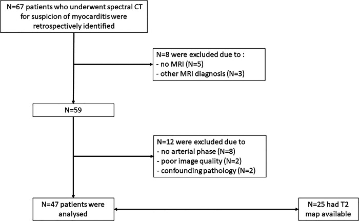
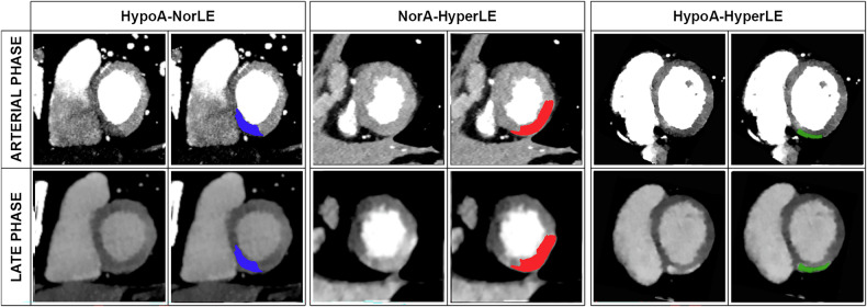
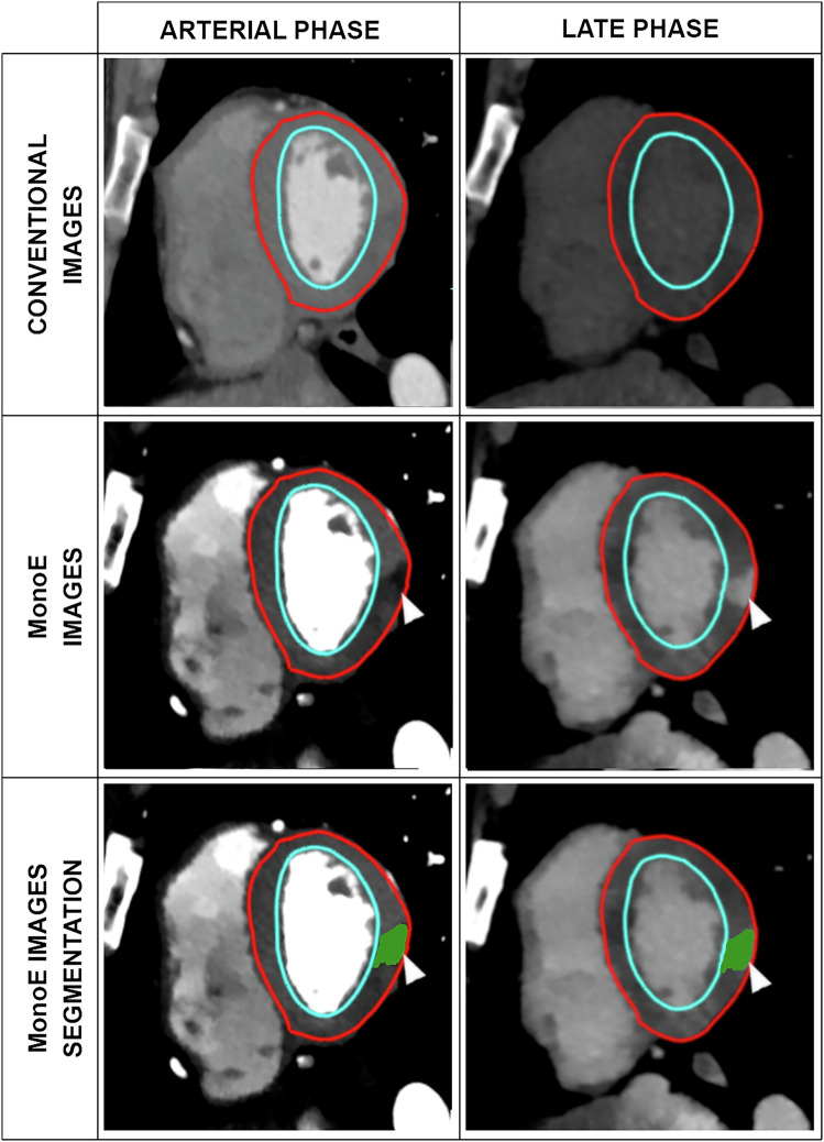
Materials and methods: Forty-seven cardiac spectral CTs performed in patients with magnetic resonance imaging (MRI)-confirmed myocarditis were retrospectively assessed. Three myocardial attenuation/enhancement patterns were visually identified and segmented on both arterial and LE acquisitions: hypodense-arterial + normal-LE (HypoArt-NorLE); normal-arterial + hyperdense-LE (NorArt-HyperLE); and hypodense-arterial + hyperdense-late (HypoArt-HyperLE). Characteristics of conventional and spectral images were calculated for all patterns and for remote myocardium. Values of HypoArt-HyperLE lesions were compared in the groups with and without edema on MRI, as assessed with T2 mapping (available for 25 patients).
Results: We found 173 lesions, 46 (26%) HypoArt-NorLE, 54 (31%) NorArt-HyperLE, and 73 (42%) HypoArt-HyperLE. On the arterial phase, HypoArt-HyperLE were more hypodense (p < 0.001) and had less iodine (0.23 mg/mL less; p < 0.001) than RM. On LE, both HypoArt-HyperLE and NorArt-HyperLE were more hyperdense and contained more iodine than the remote myocardium (all p < 0.001). HypoArt-HyperLE lesions were more hypodense and contained less iodine on the arterial phase in patients with edema on MRI as compared to those without (all p < 0.001).
Conclusion: Most myocarditis lesions detectable with spectral CT are visible on both arterial and LE acquisitions. These lesions appeared to be more pronounced on the arterial phase in patients with edema on MRI.
Relevance statement: Spectral CT arterial acquisition performed for the differential diagnosis of acute myocardial pathologies in many cases can depict myocarditis lesions as epicardial hypodense areas, most likely related to the presence of edema.
Key points: Data from spectral CT shows that most myocarditis lesions appear as hypodense on the arterial phase, matching the epicardial LE zones. A minority of myocarditis lesions appear as epicardial LE areas without anomalies of attenuation on the arterial phase. Hypodense myocardial areas are correlated to the presence of edema on MRI, suggesting they are due to the same phenomenon.

 求助内容:
求助内容: 应助结果提醒方式:


