{"title":"开发基于多模态MRI放射组学的模型来预测接受最终放疗的下咽癌患者的长期总生存率。","authors":"Xi-Wei Zhang, Dilinaer Wusiman, Ye Zhang, Xiao-Duo Yu, Su-Sheng Miao, Zhi Wang, Shao-Yan Liu, Zheng-Jiang Li, Ying Sun, Jun-Lin Yi, Chang-Ming An","doi":"10.1002/wjo2.70001","DOIUrl":null,"url":null,"abstract":"<p><strong>Objective: </strong>The aim of this study is to develop a multimodal MRI radiomics-based model for predicting long-term overall survival in hypopharyngeal cancer patients undergoing definitive radiotherapy.</p><p><strong>Methods: </strong>We enrolled 207 hypopharyngeal cancer patients who underwent definitive radiotherapy and had 5-year overall survival outcomes from two major cancer centers in China. Pretreatment MRI images and clinical features were collected. Regions of interest (ROIs) for primary tumors and lymph node metastases (LNM) were delineated on T2 and contrast-enhanced T1 (CE-T1) sequences. Principal component analysis (PCA), support vector machine (SVM), and 5-fold cross-validation were used to develop and evaluate the models.</p><p><strong>Results: </strong>Multivariate Cox regression analysis identified age under 50 years, advanced T stage, and N stage as risk factors for overall survival. Predictive models based solely on clinical features (Model A), single radiomics features (Model B), and their combination (Model C) performed poorly, with mean AUC values in the validation set of 0.663, 0.772, and 0.779, respectively. The addition of multimodal LNM and CE-T1 radiomics features significantly improved prediction accuracy (Models D and E), with AUC values of 0.831 and 0.837 in the validation set.</p><p><strong>Conclusion: </strong>We developed a well-discriminating overall survival prediction model based on multimodal MRI radiomics, applicable to patients receiving definitive radiotherapy, which may contribute to personalized treatment strategies.</p>","PeriodicalId":32097,"journal":{"name":"World Journal of OtorhinolaryngologyHead and Neck Surgery","volume":"11 3","pages":"440-448"},"PeriodicalIF":1.4000,"publicationDate":"2025-03-24","publicationTypes":"Journal Article","fieldsOfStudy":null,"isOpenAccess":false,"openAccessPdf":"https://www.ncbi.nlm.nih.gov/pmc/articles/PMC12418344/pdf/","citationCount":"0","resultStr":"{\"title\":\"Developing a multi-modal MRI radiomics-based model to predict the long-term overall survival of patients with hypopharyngeal cancer receiving definitive radiotherapy.\",\"authors\":\"Xi-Wei Zhang, Dilinaer Wusiman, Ye Zhang, Xiao-Duo Yu, Su-Sheng Miao, Zhi Wang, Shao-Yan Liu, Zheng-Jiang Li, Ying Sun, Jun-Lin Yi, Chang-Ming An\",\"doi\":\"10.1002/wjo2.70001\",\"DOIUrl\":null,\"url\":null,\"abstract\":\"<p><strong>Objective: </strong>The aim of this study is to develop a multimodal MRI radiomics-based model for predicting long-term overall survival in hypopharyngeal cancer patients undergoing definitive radiotherapy.</p><p><strong>Methods: </strong>We enrolled 207 hypopharyngeal cancer patients who underwent definitive radiotherapy and had 5-year overall survival outcomes from two major cancer centers in China. Pretreatment MRI images and clinical features were collected. Regions of interest (ROIs) for primary tumors and lymph node metastases (LNM) were delineated on T2 and contrast-enhanced T1 (CE-T1) sequences. Principal component analysis (PCA), support vector machine (SVM), and 5-fold cross-validation were used to develop and evaluate the models.</p><p><strong>Results: </strong>Multivariate Cox regression analysis identified age under 50 years, advanced T stage, and N stage as risk factors for overall survival. Predictive models based solely on clinical features (Model A), single radiomics features (Model B), and their combination (Model C) performed poorly, with mean AUC values in the validation set of 0.663, 0.772, and 0.779, respectively. The addition of multimodal LNM and CE-T1 radiomics features significantly improved prediction accuracy (Models D and E), with AUC values of 0.831 and 0.837 in the validation set.</p><p><strong>Conclusion: </strong>We developed a well-discriminating overall survival prediction model based on multimodal MRI radiomics, applicable to patients receiving definitive radiotherapy, which may contribute to personalized treatment strategies.</p>\",\"PeriodicalId\":32097,\"journal\":{\"name\":\"World Journal of OtorhinolaryngologyHead and Neck Surgery\",\"volume\":\"11 3\",\"pages\":\"440-448\"},\"PeriodicalIF\":1.4000,\"publicationDate\":\"2025-03-24\",\"publicationTypes\":\"Journal Article\",\"fieldsOfStudy\":null,\"isOpenAccess\":false,\"openAccessPdf\":\"https://www.ncbi.nlm.nih.gov/pmc/articles/PMC12418344/pdf/\",\"citationCount\":\"0\",\"resultStr\":null,\"platform\":\"Semanticscholar\",\"paperid\":null,\"PeriodicalName\":\"World Journal of OtorhinolaryngologyHead and Neck Surgery\",\"FirstCategoryId\":\"3\",\"ListUrlMain\":\"https://doi.org/10.1002/wjo2.70001\",\"RegionNum\":0,\"RegionCategory\":null,\"ArticlePicture\":[],\"TitleCN\":null,\"AbstractTextCN\":null,\"PMCID\":null,\"EPubDate\":\"2025/9/1 0:00:00\",\"PubModel\":\"eCollection\",\"JCR\":\"Q2\",\"JCRName\":\"Medicine\",\"Score\":null,\"Total\":0}","platform":"Semanticscholar","paperid":null,"PeriodicalName":"World Journal of OtorhinolaryngologyHead and Neck Surgery","FirstCategoryId":"3","ListUrlMain":"https://doi.org/10.1002/wjo2.70001","RegionNum":0,"RegionCategory":null,"ArticlePicture":[],"TitleCN":null,"AbstractTextCN":null,"PMCID":null,"EPubDate":"2025/9/1 0:00:00","PubModel":"eCollection","JCR":"Q2","JCRName":"Medicine","Score":null,"Total":0}
Developing a multi-modal MRI radiomics-based model to predict the long-term overall survival of patients with hypopharyngeal cancer receiving definitive radiotherapy.
Objective: The aim of this study is to develop a multimodal MRI radiomics-based model for predicting long-term overall survival in hypopharyngeal cancer patients undergoing definitive radiotherapy.
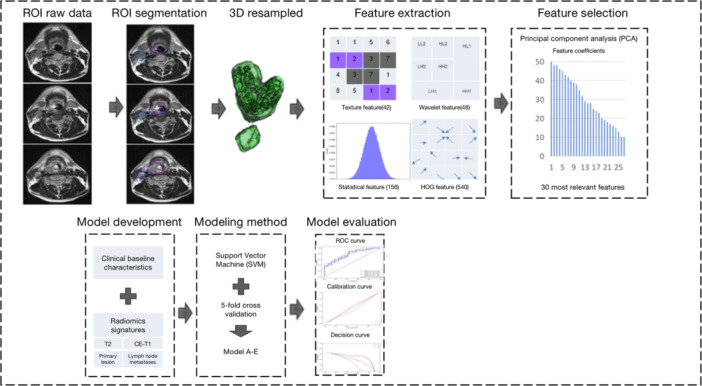
Methods: We enrolled 207 hypopharyngeal cancer patients who underwent definitive radiotherapy and had 5-year overall survival outcomes from two major cancer centers in China. Pretreatment MRI images and clinical features were collected. Regions of interest (ROIs) for primary tumors and lymph node metastases (LNM) were delineated on T2 and contrast-enhanced T1 (CE-T1) sequences. Principal component analysis (PCA), support vector machine (SVM), and 5-fold cross-validation were used to develop and evaluate the models.
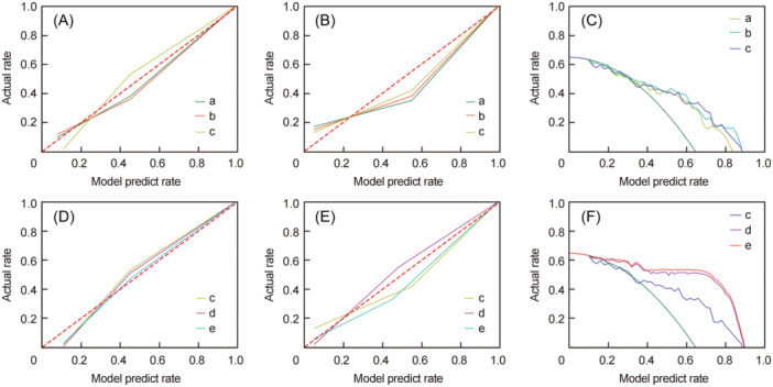
Results: Multivariate Cox regression analysis identified age under 50 years, advanced T stage, and N stage as risk factors for overall survival. Predictive models based solely on clinical features (Model A), single radiomics features (Model B), and their combination (Model C) performed poorly, with mean AUC values in the validation set of 0.663, 0.772, and 0.779, respectively. The addition of multimodal LNM and CE-T1 radiomics features significantly improved prediction accuracy (Models D and E), with AUC values of 0.831 and 0.837 in the validation set.
Conclusion: We developed a well-discriminating overall survival prediction model based on multimodal MRI radiomics, applicable to patients receiving definitive radiotherapy, which may contribute to personalized treatment strategies.

 求助内容:
求助内容: 应助结果提醒方式:


