Amandeep Singh, Manmohan Singh, Salavat R Aglyamov, David Mayerich, Kirill V Larin
{"title":"用非接触光学相干弹性成像量化骨髓弹性的年龄和空间变化。","authors":"Amandeep Singh, Manmohan Singh, Salavat R Aglyamov, David Mayerich, Kirill V Larin","doi":"10.1117/1.JBO.30.12.124505","DOIUrl":null,"url":null,"abstract":"<p><strong>Significance: </strong>The bone marrow is essential in immune regulation to maintain body homeostasis and to control the trafficking of stromal cells. A framework of connective tissue upholds bone marrow cells to maintain their mechanical and functional integrity. The biomechanical characterization of the bone marrow may provide useful insights for diagnosing hematologic diseases such as primary myelofibrosis. Optical coherence elastography (OCE) can measure the mechanical properties of tissues with high spatiotemporal resolution and may be well-suited for characterizing bone marrow elasticity.</p><p><strong>Aim: </strong>We demonstrate the quantification of the elastic modulus of bone marrow <i>ex vivo</i> at different locations along the diaphysis of mice femurs and compare the elastic modulus within different age groups of mice femurs.</p><p><strong>Approach: </strong>The femur bone marrow of CD1 mice, <math><mrow><mo>∼</mo> <mn>12</mn></mrow> </math> weeks old (young adult), 24 weeks old (mature adult), and 1 year old (old adult), was imaged with OCE ( <math><mrow><mi>N</mi> <mo>=</mo> <mn>4</mn></mrow> </math> femurs for each age group) to investigate the change in stiffness with age and location along the femur. A noncontact air-coupled ultrasound (ACUS) transducer induced elastic waves in the bone marrow, which were detected by phase-sensitive optical coherence tomography. The ACUS-OCE measurements were taken at three different locations along the diaphysis from the proximal end to the distal end to investigate the spatial stiffness variations.</p><p><strong>Results: </strong>The results show that the stiffness of femoral bone marrow increases significantly with age ( <math><mrow><mi>p</mi> <mo><</mo> <mn>0.001</mn></mrow> </math> ), but there was no significant difference in Young's moduli among the locations for young ( <math> <mrow> <msup><mrow><mi>χ</mi></mrow> <mrow><mn>2</mn></mrow> </msup> <mo>(</mo> <mn>2</mn> <mo>)</mo> <mo>=</mo> <mn>2.15</mn></mrow> </math> , <math><mrow><mi>p</mi> <mo>=</mo> <mn>0.33</mn></mrow> </math> ), mature ( <math> <mrow> <msup><mrow><mi>χ</mi></mrow> <mrow><mn>2</mn></mrow> </msup> <mo>(</mo> <mn>2</mn> <mo>)</mo> <mo>=</mo> <mn>5.68</mn></mrow> </math> , <math><mrow><mi>p</mi> <mo>=</mo> <mn>0.058</mn></mrow> </math> ), and old ( <math> <mrow> <msup><mrow><mi>χ</mi></mrow> <mrow><mn>2</mn></mrow> </msup> <mo>(</mo> <mn>2</mn> <mo>)</mo> <mo>=</mo> <mn>5.73</mn></mrow> </math> , <math><mrow><mi>p</mi> <mo>=</mo> <mn>0.056</mn></mrow> </math> ) mice femur samples.</p><p><strong>Conclusions: </strong>These findings show that OCE is promising for mapping the stiffness of the intact bone marrow and could be used for minimally invasive clinical applications.</p>","PeriodicalId":15264,"journal":{"name":"Journal of Biomedical Optics","volume":"30 12","pages":"124505"},"PeriodicalIF":2.9000,"publicationDate":"2025-12-01","publicationTypes":"Journal Article","fieldsOfStudy":null,"isOpenAccess":false,"openAccessPdf":"https://www.ncbi.nlm.nih.gov/pmc/articles/PMC12422286/pdf/","citationCount":"0","resultStr":"{\"title\":\"Quantifying age and spatial variations of bone marrow elasticity with noncontact optical coherence elastography.\",\"authors\":\"Amandeep Singh, Manmohan Singh, Salavat R Aglyamov, David Mayerich, Kirill V Larin\",\"doi\":\"10.1117/1.JBO.30.12.124505\",\"DOIUrl\":null,\"url\":null,\"abstract\":\"<p><strong>Significance: </strong>The bone marrow is essential in immune regulation to maintain body homeostasis and to control the trafficking of stromal cells. A framework of connective tissue upholds bone marrow cells to maintain their mechanical and functional integrity. The biomechanical characterization of the bone marrow may provide useful insights for diagnosing hematologic diseases such as primary myelofibrosis. Optical coherence elastography (OCE) can measure the mechanical properties of tissues with high spatiotemporal resolution and may be well-suited for characterizing bone marrow elasticity.</p><p><strong>Aim: </strong>We demonstrate the quantification of the elastic modulus of bone marrow <i>ex vivo</i> at different locations along the diaphysis of mice femurs and compare the elastic modulus within different age groups of mice femurs.</p><p><strong>Approach: </strong>The femur bone marrow of CD1 mice, <math><mrow><mo>∼</mo> <mn>12</mn></mrow> </math> weeks old (young adult), 24 weeks old (mature adult), and 1 year old (old adult), was imaged with OCE ( <math><mrow><mi>N</mi> <mo>=</mo> <mn>4</mn></mrow> </math> femurs for each age group) to investigate the change in stiffness with age and location along the femur. A noncontact air-coupled ultrasound (ACUS) transducer induced elastic waves in the bone marrow, which were detected by phase-sensitive optical coherence tomography. The ACUS-OCE measurements were taken at three different locations along the diaphysis from the proximal end to the distal end to investigate the spatial stiffness variations.</p><p><strong>Results: </strong>The results show that the stiffness of femoral bone marrow increases significantly with age ( <math><mrow><mi>p</mi> <mo><</mo> <mn>0.001</mn></mrow> </math> ), but there was no significant difference in Young's moduli among the locations for young ( <math> <mrow> <msup><mrow><mi>χ</mi></mrow> <mrow><mn>2</mn></mrow> </msup> <mo>(</mo> <mn>2</mn> <mo>)</mo> <mo>=</mo> <mn>2.15</mn></mrow> </math> , <math><mrow><mi>p</mi> <mo>=</mo> <mn>0.33</mn></mrow> </math> ), mature ( <math> <mrow> <msup><mrow><mi>χ</mi></mrow> <mrow><mn>2</mn></mrow> </msup> <mo>(</mo> <mn>2</mn> <mo>)</mo> <mo>=</mo> <mn>5.68</mn></mrow> </math> , <math><mrow><mi>p</mi> <mo>=</mo> <mn>0.058</mn></mrow> </math> ), and old ( <math> <mrow> <msup><mrow><mi>χ</mi></mrow> <mrow><mn>2</mn></mrow> </msup> <mo>(</mo> <mn>2</mn> <mo>)</mo> <mo>=</mo> <mn>5.73</mn></mrow> </math> , <math><mrow><mi>p</mi> <mo>=</mo> <mn>0.056</mn></mrow> </math> ) mice femur samples.</p><p><strong>Conclusions: </strong>These findings show that OCE is promising for mapping the stiffness of the intact bone marrow and could be used for minimally invasive clinical applications.</p>\",\"PeriodicalId\":15264,\"journal\":{\"name\":\"Journal of Biomedical Optics\",\"volume\":\"30 12\",\"pages\":\"124505\"},\"PeriodicalIF\":2.9000,\"publicationDate\":\"2025-12-01\",\"publicationTypes\":\"Journal Article\",\"fieldsOfStudy\":null,\"isOpenAccess\":false,\"openAccessPdf\":\"https://www.ncbi.nlm.nih.gov/pmc/articles/PMC12422286/pdf/\",\"citationCount\":\"0\",\"resultStr\":null,\"platform\":\"Semanticscholar\",\"paperid\":null,\"PeriodicalName\":\"Journal of Biomedical Optics\",\"FirstCategoryId\":\"3\",\"ListUrlMain\":\"https://doi.org/10.1117/1.JBO.30.12.124505\",\"RegionNum\":3,\"RegionCategory\":\"医学\",\"ArticlePicture\":[],\"TitleCN\":null,\"AbstractTextCN\":null,\"PMCID\":null,\"EPubDate\":\"2025/9/10 0:00:00\",\"PubModel\":\"Epub\",\"JCR\":\"Q2\",\"JCRName\":\"BIOCHEMICAL RESEARCH METHODS\",\"Score\":null,\"Total\":0}","platform":"Semanticscholar","paperid":null,"PeriodicalName":"Journal of Biomedical Optics","FirstCategoryId":"3","ListUrlMain":"https://doi.org/10.1117/1.JBO.30.12.124505","RegionNum":3,"RegionCategory":"医学","ArticlePicture":[],"TitleCN":null,"AbstractTextCN":null,"PMCID":null,"EPubDate":"2025/9/10 0:00:00","PubModel":"Epub","JCR":"Q2","JCRName":"BIOCHEMICAL RESEARCH METHODS","Score":null,"Total":0}
引用次数: 0
摘要
意义:骨髓在维持机体稳态和控制基质细胞运输的免疫调节中起着至关重要的作用。结缔组织的框架支撑着骨髓细胞,以维持其机械和功能的完整性。骨髓的生物力学特征可能为诊断血液病(如原发性骨髓纤维化)提供有用的见解。光学相干弹性成像(OCE)可以以高时空分辨率测量组织的力学特性,可能非常适合表征骨髓弹性。目的:定量测定小鼠股骨骨干不同部位离体骨髓弹性模量,并比较不同年龄组小鼠股骨弹性模量。方法:对CD1小鼠,12周龄(青壮年),24周龄(成年)和1岁(老年)的股骨骨髓进行OCE成像(每个年龄组N = 4根股骨),以研究股骨沿年龄和位置的刚度变化。非接触式空气耦合超声(ACUS)换能器在骨髓中产生弹性波,用相敏光学相干层析成像技术检测弹性波。ACUS-OCE测量从近端到远端沿骨干的三个不同位置进行,以研究空间刚度变化。结果:小鼠股骨骨髓刚度随年龄的增长而显著增加(p < 0.001),但幼鼠(χ 2 (2) = 2.15, p = 0.33)、成年鼠(χ 2 (2) = 5.68, p = 0.058)、老年鼠(χ 2 (2) = 5.73, p = 0.056)股骨样本的杨氏模量在不同部位无显著差异。结论:这些发现表明OCE有希望绘制完整骨髓的硬度,并可用于微创临床应用。
Quantifying age and spatial variations of bone marrow elasticity with noncontact optical coherence elastography.
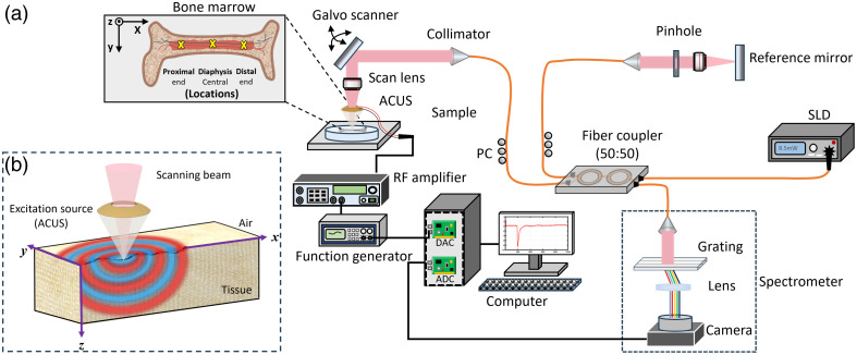
Significance: The bone marrow is essential in immune regulation to maintain body homeostasis and to control the trafficking of stromal cells. A framework of connective tissue upholds bone marrow cells to maintain their mechanical and functional integrity. The biomechanical characterization of the bone marrow may provide useful insights for diagnosing hematologic diseases such as primary myelofibrosis. Optical coherence elastography (OCE) can measure the mechanical properties of tissues with high spatiotemporal resolution and may be well-suited for characterizing bone marrow elasticity.
Aim: We demonstrate the quantification of the elastic modulus of bone marrow ex vivo at different locations along the diaphysis of mice femurs and compare the elastic modulus within different age groups of mice femurs.
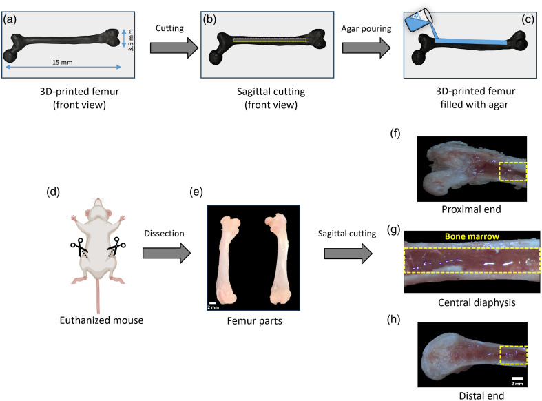
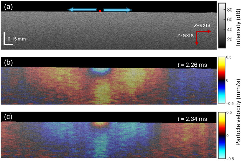
Approach: The femur bone marrow of CD1 mice, weeks old (young adult), 24 weeks old (mature adult), and 1 year old (old adult), was imaged with OCE ( femurs for each age group) to investigate the change in stiffness with age and location along the femur. A noncontact air-coupled ultrasound (ACUS) transducer induced elastic waves in the bone marrow, which were detected by phase-sensitive optical coherence tomography. The ACUS-OCE measurements were taken at three different locations along the diaphysis from the proximal end to the distal end to investigate the spatial stiffness variations.
Results: The results show that the stiffness of femoral bone marrow increases significantly with age ( ), but there was no significant difference in Young's moduli among the locations for young ( , ), mature ( , ), and old ( , ) mice femur samples.
Conclusions: These findings show that OCE is promising for mapping the stiffness of the intact bone marrow and could be used for minimally invasive clinical applications.
期刊介绍:
The Journal of Biomedical Optics publishes peer-reviewed papers on the use of modern optical technology for improved health care and biomedical research.

 求助内容:
求助内容: 应助结果提醒方式:


