Garima Anandani, Anita Motiani, Parth Goswami, Amit Sonagra
{"title":"与利用高效液相色谱仪技术识别异常血红蛋白变异相关的挑战:古吉拉特邦一家医院的前瞻性研究。","authors":"Garima Anandani, Anita Motiani, Parth Goswami, Amit Sonagra","doi":"10.4103/ijabmr.ijabmr_70_25","DOIUrl":null,"url":null,"abstract":"<p><strong>Introduction: </strong>Cation exchange high-performance liquid chromatography (HPLC) serves as a rapid, reproducible, and accurate method for diagnosing hemoglobinopathies. This study outlines the diagnostic approach and the challenges faced in the routine diagnosis of hemoglobinopathies through HPLC, particularly in laboratories with limited resources.</p><p><strong>Aims and objectives: </strong>The aim of the study was to identify the challenges encountered in identifying abnormal hemoglobin (Hb) variants and to determine the significance of HbA2 and/or fetal Hb (HbF) analysis in the HPLC methodology for hemoglobinopathies.</p><p><strong>Materials and methods: </strong>A total of 1900 samples were analyzed using the ARKRAY ADAMS HA-8180T HPLC automated analyzer for the purpose of hemoglobinopathy testing. The samples were classified into normal or abnormal hemoglobin variants based on the percentage levels of HbA2, HbF, HbA, and the identification of any abnormal peaks. Among these, 113 cases were diagnosed to have thalassemia or hemoglobinopathy. The clinical presentations and red blood cell (RBC) indices were compared with the HPLC findings for each case, thereby contributing to the accuracy of the diagnosis.</p><p><strong>Results: </strong>The study examined the distribution of Hb variants, revealing that β-thalassemia trait was the most prevalent at 44.2%, followed by sickle cell trait at 13.3% and HbD Punjab trait at 10.6%. There were many challenging cases with elevated HbA2, like HbE thalassemia and Hb Lepore. Furthermore, there was identification of some abnormal peaks which were not exactly in the instrument's predetermined HbA2, HbF, HbA, or sickle windows, like HbJ Meerut. There were a few cases with abnormally elevated HbF, which can be seen in homozygous β-thalassemia, sickle cell disease, compound double heterozygous sickle cell β-thalassemia, δβ-thalassemia, and hereditary persistence of HbF. Carriers of β-thalassemia were generally identified by an HbA2 level of 4% or higher; however, there were nine cases which exhibited borderline HbA2 levels ranging from 3.5% to 3.9%, which might turn out to be β-thalassemia trait, especially in high-prevalence areas like Gujarat.</p><p><strong>Conclusion: </strong>Any case scenario with abnormally elevated HbA2 is not always β-thalassemia trait. Nor abnormally elevated HbF may always indicate β-thalassemia major. Furthermore, some clinico-pathologically relevant hemoglobinopathies might show an abnormal peak on HPLC at any retention time, which may not be necessarily determined by the machine to be in some specific window. We need to correlate the clinical context, RBC indices, HPLC findings, and family studies to effectively detect most Hb variants.</p>","PeriodicalId":13727,"journal":{"name":"International Journal of Applied and Basic Medical Research","volume":"15 3","pages":"197-205"},"PeriodicalIF":0.8000,"publicationDate":"2025-07-01","publicationTypes":"Journal Article","fieldsOfStudy":null,"isOpenAccess":false,"openAccessPdf":"https://www.ncbi.nlm.nih.gov/pmc/articles/PMC12422550/pdf/","citationCount":"0","resultStr":"{\"title\":\"Challenges Associated with the Identification of Abnormal Hemoglobin Variants Utilizing the High-performance Liquid Chromatograph Technique: A Prospective Study in a Hospital Setting in Gujarat.\",\"authors\":\"Garima Anandani, Anita Motiani, Parth Goswami, Amit Sonagra\",\"doi\":\"10.4103/ijabmr.ijabmr_70_25\",\"DOIUrl\":null,\"url\":null,\"abstract\":\"<p><strong>Introduction: </strong>Cation exchange high-performance liquid chromatography (HPLC) serves as a rapid, reproducible, and accurate method for diagnosing hemoglobinopathies. This study outlines the diagnostic approach and the challenges faced in the routine diagnosis of hemoglobinopathies through HPLC, particularly in laboratories with limited resources.</p><p><strong>Aims and objectives: </strong>The aim of the study was to identify the challenges encountered in identifying abnormal hemoglobin (Hb) variants and to determine the significance of HbA2 and/or fetal Hb (HbF) analysis in the HPLC methodology for hemoglobinopathies.</p><p><strong>Materials and methods: </strong>A total of 1900 samples were analyzed using the ARKRAY ADAMS HA-8180T HPLC automated analyzer for the purpose of hemoglobinopathy testing. The samples were classified into normal or abnormal hemoglobin variants based on the percentage levels of HbA2, HbF, HbA, and the identification of any abnormal peaks. Among these, 113 cases were diagnosed to have thalassemia or hemoglobinopathy. The clinical presentations and red blood cell (RBC) indices were compared with the HPLC findings for each case, thereby contributing to the accuracy of the diagnosis.</p><p><strong>Results: </strong>The study examined the distribution of Hb variants, revealing that β-thalassemia trait was the most prevalent at 44.2%, followed by sickle cell trait at 13.3% and HbD Punjab trait at 10.6%. There were many challenging cases with elevated HbA2, like HbE thalassemia and Hb Lepore. Furthermore, there was identification of some abnormal peaks which were not exactly in the instrument's predetermined HbA2, HbF, HbA, or sickle windows, like HbJ Meerut. There were a few cases with abnormally elevated HbF, which can be seen in homozygous β-thalassemia, sickle cell disease, compound double heterozygous sickle cell β-thalassemia, δβ-thalassemia, and hereditary persistence of HbF. Carriers of β-thalassemia were generally identified by an HbA2 level of 4% or higher; however, there were nine cases which exhibited borderline HbA2 levels ranging from 3.5% to 3.9%, which might turn out to be β-thalassemia trait, especially in high-prevalence areas like Gujarat.</p><p><strong>Conclusion: </strong>Any case scenario with abnormally elevated HbA2 is not always β-thalassemia trait. Nor abnormally elevated HbF may always indicate β-thalassemia major. Furthermore, some clinico-pathologically relevant hemoglobinopathies might show an abnormal peak on HPLC at any retention time, which may not be necessarily determined by the machine to be in some specific window. We need to correlate the clinical context, RBC indices, HPLC findings, and family studies to effectively detect most Hb variants.</p>\",\"PeriodicalId\":13727,\"journal\":{\"name\":\"International Journal of Applied and Basic Medical Research\",\"volume\":\"15 3\",\"pages\":\"197-205\"},\"PeriodicalIF\":0.8000,\"publicationDate\":\"2025-07-01\",\"publicationTypes\":\"Journal Article\",\"fieldsOfStudy\":null,\"isOpenAccess\":false,\"openAccessPdf\":\"https://www.ncbi.nlm.nih.gov/pmc/articles/PMC12422550/pdf/\",\"citationCount\":\"0\",\"resultStr\":null,\"platform\":\"Semanticscholar\",\"paperid\":null,\"PeriodicalName\":\"International Journal of Applied and Basic Medical Research\",\"FirstCategoryId\":\"1085\",\"ListUrlMain\":\"https://doi.org/10.4103/ijabmr.ijabmr_70_25\",\"RegionNum\":0,\"RegionCategory\":null,\"ArticlePicture\":[],\"TitleCN\":null,\"AbstractTextCN\":null,\"PMCID\":null,\"EPubDate\":\"2025/8/20 0:00:00\",\"PubModel\":\"Epub\",\"JCR\":\"Q3\",\"JCRName\":\"MEDICINE, GENERAL & INTERNAL\",\"Score\":null,\"Total\":0}","platform":"Semanticscholar","paperid":null,"PeriodicalName":"International Journal of Applied and Basic Medical Research","FirstCategoryId":"1085","ListUrlMain":"https://doi.org/10.4103/ijabmr.ijabmr_70_25","RegionNum":0,"RegionCategory":null,"ArticlePicture":[],"TitleCN":null,"AbstractTextCN":null,"PMCID":null,"EPubDate":"2025/8/20 0:00:00","PubModel":"Epub","JCR":"Q3","JCRName":"MEDICINE, GENERAL & INTERNAL","Score":null,"Total":0}
Challenges Associated with the Identification of Abnormal Hemoglobin Variants Utilizing the High-performance Liquid Chromatograph Technique: A Prospective Study in a Hospital Setting in Gujarat.
Introduction: Cation exchange high-performance liquid chromatography (HPLC) serves as a rapid, reproducible, and accurate method for diagnosing hemoglobinopathies. This study outlines the diagnostic approach and the challenges faced in the routine diagnosis of hemoglobinopathies through HPLC, particularly in laboratories with limited resources.
Aims and objectives: The aim of the study was to identify the challenges encountered in identifying abnormal hemoglobin (Hb) variants and to determine the significance of HbA2 and/or fetal Hb (HbF) analysis in the HPLC methodology for hemoglobinopathies.
Materials and methods: A total of 1900 samples were analyzed using the ARKRAY ADAMS HA-8180T HPLC automated analyzer for the purpose of hemoglobinopathy testing. The samples were classified into normal or abnormal hemoglobin variants based on the percentage levels of HbA2, HbF, HbA, and the identification of any abnormal peaks. Among these, 113 cases were diagnosed to have thalassemia or hemoglobinopathy. The clinical presentations and red blood cell (RBC) indices were compared with the HPLC findings for each case, thereby contributing to the accuracy of the diagnosis.
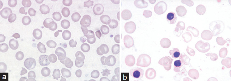
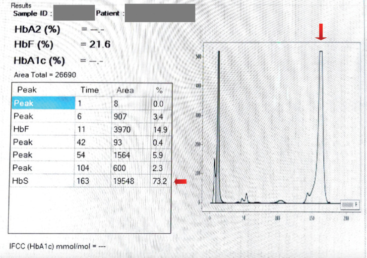
Results: The study examined the distribution of Hb variants, revealing that β-thalassemia trait was the most prevalent at 44.2%, followed by sickle cell trait at 13.3% and HbD Punjab trait at 10.6%. There were many challenging cases with elevated HbA2, like HbE thalassemia and Hb Lepore. Furthermore, there was identification of some abnormal peaks which were not exactly in the instrument's predetermined HbA2, HbF, HbA, or sickle windows, like HbJ Meerut. There were a few cases with abnormally elevated HbF, which can be seen in homozygous β-thalassemia, sickle cell disease, compound double heterozygous sickle cell β-thalassemia, δβ-thalassemia, and hereditary persistence of HbF. Carriers of β-thalassemia were generally identified by an HbA2 level of 4% or higher; however, there were nine cases which exhibited borderline HbA2 levels ranging from 3.5% to 3.9%, which might turn out to be β-thalassemia trait, especially in high-prevalence areas like Gujarat.
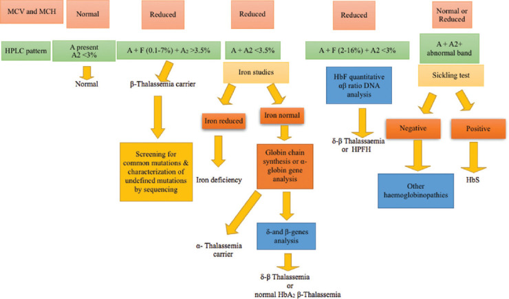
Conclusion: Any case scenario with abnormally elevated HbA2 is not always β-thalassemia trait. Nor abnormally elevated HbF may always indicate β-thalassemia major. Furthermore, some clinico-pathologically relevant hemoglobinopathies might show an abnormal peak on HPLC at any retention time, which may not be necessarily determined by the machine to be in some specific window. We need to correlate the clinical context, RBC indices, HPLC findings, and family studies to effectively detect most Hb variants.

 求助内容:
求助内容: 应助结果提醒方式:


