{"title":"心脏磁共振作为发现异常弥散性组织胞浆菌病的关键:1例报告。","authors":"Giorgia Benzoni, Ilaria Garofani, Diana Artioli, Cristina Giannattasio, Patrizia Pedrotti","doi":"10.1093/ehjcr/ytaf408","DOIUrl":null,"url":null,"abstract":"<p><strong>Background: </strong>Disseminated histoplasmosis is a severe fungal infection caused by <i>Histoplasma capsulatum</i> which primarily affects immunocompromised individuals, leading to widespread infection in multiple organs such as lungs, liver, and spleen. Early diagnosis and treatment are crucial for effective management.</p><p><strong>Case summary: </strong>We herein report the case of a 33-year-old male patient who presented to the Emergency Department with fever and chest pain after returning from a journey from a tropical region in Centre America. Initial tests showed elevated high-sensitivity troponin T (Hs-TnT) levels, suggesting possible cardiac involvement, but EKG and chest X-ray were normal. Echocardiography detected hypokinesis of the interventricular septum and a small pericardial effusion. Cardiac magnetic resonance (CMR) showed left ventricular function at lower normal limits and a small pericardial effusion, but also masses in the lungs and mediastinum, confirmed by computed tomography. Biopsy was performed, and histology revealed disseminated histoplasmosis. The patient was treated with antifungals and was discharged after two weeks, continuing antifungal administration in the outpatient clinic for 18 months. Follow-up imaging showed significant reduction of the masses. The patient remained asymptomatic with no further treatment needed.</p><p><strong>Discussion: </strong>In this case report, we emphasize the essential role of a multimodal imaging approach in diagnosing cardiac inflammatory diseases. CMR was pivotal providing a three-dimensional perspective of the mediastinum, which led to the identification of a retrocardiac mediastinal mass that might have otherwise gone undetected. This highlights the importance of integrating multimodality imaging techniques to improve diagnostic accuracy and guide effective treatment strategies.</p>","PeriodicalId":11910,"journal":{"name":"European Heart Journal: Case Reports","volume":"9 9","pages":"ytaf408"},"PeriodicalIF":0.8000,"publicationDate":"2025-08-21","publicationTypes":"Journal Article","fieldsOfStudy":null,"isOpenAccess":false,"openAccessPdf":"https://www.ncbi.nlm.nih.gov/pmc/articles/PMC12418945/pdf/","citationCount":"0","resultStr":"{\"title\":\"Cardiac magnetic resonance as the key to uncovering unusual disseminated histoplasmosis: a case report.\",\"authors\":\"Giorgia Benzoni, Ilaria Garofani, Diana Artioli, Cristina Giannattasio, Patrizia Pedrotti\",\"doi\":\"10.1093/ehjcr/ytaf408\",\"DOIUrl\":null,\"url\":null,\"abstract\":\"<p><strong>Background: </strong>Disseminated histoplasmosis is a severe fungal infection caused by <i>Histoplasma capsulatum</i> which primarily affects immunocompromised individuals, leading to widespread infection in multiple organs such as lungs, liver, and spleen. Early diagnosis and treatment are crucial for effective management.</p><p><strong>Case summary: </strong>We herein report the case of a 33-year-old male patient who presented to the Emergency Department with fever and chest pain after returning from a journey from a tropical region in Centre America. Initial tests showed elevated high-sensitivity troponin T (Hs-TnT) levels, suggesting possible cardiac involvement, but EKG and chest X-ray were normal. Echocardiography detected hypokinesis of the interventricular septum and a small pericardial effusion. Cardiac magnetic resonance (CMR) showed left ventricular function at lower normal limits and a small pericardial effusion, but also masses in the lungs and mediastinum, confirmed by computed tomography. Biopsy was performed, and histology revealed disseminated histoplasmosis. The patient was treated with antifungals and was discharged after two weeks, continuing antifungal administration in the outpatient clinic for 18 months. Follow-up imaging showed significant reduction of the masses. The patient remained asymptomatic with no further treatment needed.</p><p><strong>Discussion: </strong>In this case report, we emphasize the essential role of a multimodal imaging approach in diagnosing cardiac inflammatory diseases. CMR was pivotal providing a three-dimensional perspective of the mediastinum, which led to the identification of a retrocardiac mediastinal mass that might have otherwise gone undetected. This highlights the importance of integrating multimodality imaging techniques to improve diagnostic accuracy and guide effective treatment strategies.</p>\",\"PeriodicalId\":11910,\"journal\":{\"name\":\"European Heart Journal: Case Reports\",\"volume\":\"9 9\",\"pages\":\"ytaf408\"},\"PeriodicalIF\":0.8000,\"publicationDate\":\"2025-08-21\",\"publicationTypes\":\"Journal Article\",\"fieldsOfStudy\":null,\"isOpenAccess\":false,\"openAccessPdf\":\"https://www.ncbi.nlm.nih.gov/pmc/articles/PMC12418945/pdf/\",\"citationCount\":\"0\",\"resultStr\":null,\"platform\":\"Semanticscholar\",\"paperid\":null,\"PeriodicalName\":\"European Heart Journal: Case Reports\",\"FirstCategoryId\":\"1085\",\"ListUrlMain\":\"https://doi.org/10.1093/ehjcr/ytaf408\",\"RegionNum\":0,\"RegionCategory\":null,\"ArticlePicture\":[],\"TitleCN\":null,\"AbstractTextCN\":null,\"PMCID\":null,\"EPubDate\":\"2025/9/1 0:00:00\",\"PubModel\":\"eCollection\",\"JCR\":\"Q4\",\"JCRName\":\"CARDIAC & CARDIOVASCULAR SYSTEMS\",\"Score\":null,\"Total\":0}","platform":"Semanticscholar","paperid":null,"PeriodicalName":"European Heart Journal: Case Reports","FirstCategoryId":"1085","ListUrlMain":"https://doi.org/10.1093/ehjcr/ytaf408","RegionNum":0,"RegionCategory":null,"ArticlePicture":[],"TitleCN":null,"AbstractTextCN":null,"PMCID":null,"EPubDate":"2025/9/1 0:00:00","PubModel":"eCollection","JCR":"Q4","JCRName":"CARDIAC & CARDIOVASCULAR SYSTEMS","Score":null,"Total":0}
Cardiac magnetic resonance as the key to uncovering unusual disseminated histoplasmosis: a case report.
Background: Disseminated histoplasmosis is a severe fungal infection caused by Histoplasma capsulatum which primarily affects immunocompromised individuals, leading to widespread infection in multiple organs such as lungs, liver, and spleen. Early diagnosis and treatment are crucial for effective management.
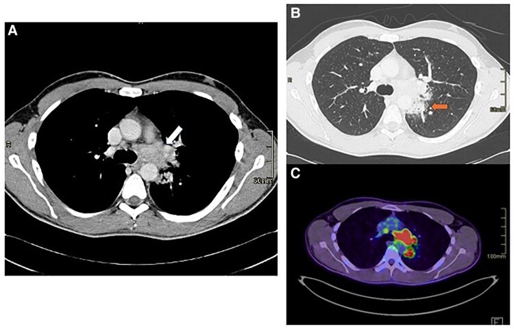
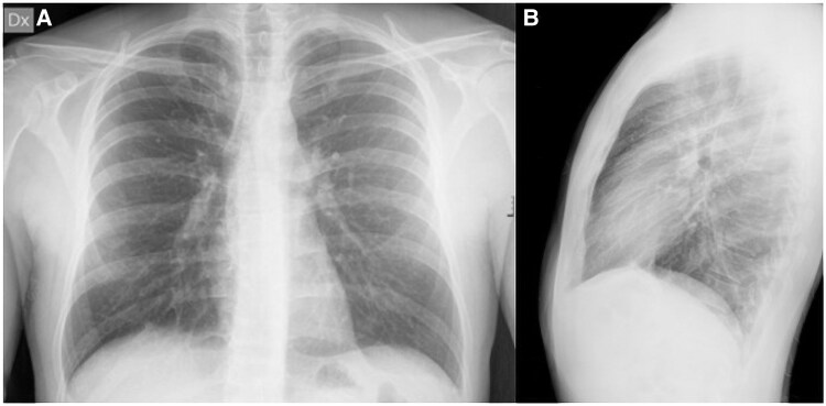
Case summary: We herein report the case of a 33-year-old male patient who presented to the Emergency Department with fever and chest pain after returning from a journey from a tropical region in Centre America. Initial tests showed elevated high-sensitivity troponin T (Hs-TnT) levels, suggesting possible cardiac involvement, but EKG and chest X-ray were normal. Echocardiography detected hypokinesis of the interventricular septum and a small pericardial effusion. Cardiac magnetic resonance (CMR) showed left ventricular function at lower normal limits and a small pericardial effusion, but also masses in the lungs and mediastinum, confirmed by computed tomography. Biopsy was performed, and histology revealed disseminated histoplasmosis. The patient was treated with antifungals and was discharged after two weeks, continuing antifungal administration in the outpatient clinic for 18 months. Follow-up imaging showed significant reduction of the masses. The patient remained asymptomatic with no further treatment needed.
Discussion: In this case report, we emphasize the essential role of a multimodal imaging approach in diagnosing cardiac inflammatory diseases. CMR was pivotal providing a three-dimensional perspective of the mediastinum, which led to the identification of a retrocardiac mediastinal mass that might have otherwise gone undetected. This highlights the importance of integrating multimodality imaging techniques to improve diagnostic accuracy and guide effective treatment strategies.


 求助内容:
求助内容: 应助结果提醒方式:


