José Martín Alanís-Naranjo, Omar Alejandro Gil-Guzmán, Daniel Velasco-Ortiz, Ma Delia Pérez Montiel-Gómez
{"title":"孤立性纤维性肿瘤心包内定位1例。","authors":"José Martín Alanís-Naranjo, Omar Alejandro Gil-Guzmán, Daniel Velasco-Ortiz, Ma Delia Pérez Montiel-Gómez","doi":"10.1093/ehjcr/ytaf422","DOIUrl":null,"url":null,"abstract":"<p><strong>Background: </strong>A solitary fibrous tumour (SFT) is a rare fibroblastic tumour, with primary cardiac SFT extremely rare. We report a rare case of a patient presenting with sudden dyspnoea who was diagnosed with a primary cardiac SFT.</p><p><strong>Case summary: </strong>A 56-year-old woman with a history of diabetes and active smoking presented with sudden dyspnoea. Computed tomography scan revealed a mass adjacent to the left ventricle. In addition to the large pericardial effusion, no signs of cardiac tamponade or valvulopathies were found on echocardiography. The patient underwent open-heart surgery and mass removal, finding a tumour located inside the pericardial sac and attached to the left ventricle's lateral wall; it did not invade other heart structures. Histological and immunohistochemical examination of the mass revealed SFT diagnosis. The patient was discharged from the hospital in full health, and follow-up examinations revealed no evidence of tumour recurrence.</p><p><strong>Discussion: </strong>Solitary fibrous tumour most commonly occurs in middle-aged patients and is not gender specific. Multimodal imaging is crucial for diagnosing and managing SFT. A definitive diagnosis must be based on both immunohistochemical and histopathological findings. STAT6 immunoexpression is the most reliable marker for histopathology diagnosis. Given the high SFT recurrence rate, follow-up is essential.</p>","PeriodicalId":11910,"journal":{"name":"European Heart Journal: Case Reports","volume":"9 9","pages":"ytaf422"},"PeriodicalIF":0.8000,"publicationDate":"2025-08-26","publicationTypes":"Journal Article","fieldsOfStudy":null,"isOpenAccess":false,"openAccessPdf":"https://www.ncbi.nlm.nih.gov/pmc/articles/PMC12418934/pdf/","citationCount":"0","resultStr":"{\"title\":\"Intrapericardial localization of solitary fibrous tumour: a case report.\",\"authors\":\"José Martín Alanís-Naranjo, Omar Alejandro Gil-Guzmán, Daniel Velasco-Ortiz, Ma Delia Pérez Montiel-Gómez\",\"doi\":\"10.1093/ehjcr/ytaf422\",\"DOIUrl\":null,\"url\":null,\"abstract\":\"<p><strong>Background: </strong>A solitary fibrous tumour (SFT) is a rare fibroblastic tumour, with primary cardiac SFT extremely rare. We report a rare case of a patient presenting with sudden dyspnoea who was diagnosed with a primary cardiac SFT.</p><p><strong>Case summary: </strong>A 56-year-old woman with a history of diabetes and active smoking presented with sudden dyspnoea. Computed tomography scan revealed a mass adjacent to the left ventricle. In addition to the large pericardial effusion, no signs of cardiac tamponade or valvulopathies were found on echocardiography. The patient underwent open-heart surgery and mass removal, finding a tumour located inside the pericardial sac and attached to the left ventricle's lateral wall; it did not invade other heart structures. Histological and immunohistochemical examination of the mass revealed SFT diagnosis. The patient was discharged from the hospital in full health, and follow-up examinations revealed no evidence of tumour recurrence.</p><p><strong>Discussion: </strong>Solitary fibrous tumour most commonly occurs in middle-aged patients and is not gender specific. Multimodal imaging is crucial for diagnosing and managing SFT. A definitive diagnosis must be based on both immunohistochemical and histopathological findings. STAT6 immunoexpression is the most reliable marker for histopathology diagnosis. Given the high SFT recurrence rate, follow-up is essential.</p>\",\"PeriodicalId\":11910,\"journal\":{\"name\":\"European Heart Journal: Case Reports\",\"volume\":\"9 9\",\"pages\":\"ytaf422\"},\"PeriodicalIF\":0.8000,\"publicationDate\":\"2025-08-26\",\"publicationTypes\":\"Journal Article\",\"fieldsOfStudy\":null,\"isOpenAccess\":false,\"openAccessPdf\":\"https://www.ncbi.nlm.nih.gov/pmc/articles/PMC12418934/pdf/\",\"citationCount\":\"0\",\"resultStr\":null,\"platform\":\"Semanticscholar\",\"paperid\":null,\"PeriodicalName\":\"European Heart Journal: Case Reports\",\"FirstCategoryId\":\"1085\",\"ListUrlMain\":\"https://doi.org/10.1093/ehjcr/ytaf422\",\"RegionNum\":0,\"RegionCategory\":null,\"ArticlePicture\":[],\"TitleCN\":null,\"AbstractTextCN\":null,\"PMCID\":null,\"EPubDate\":\"2025/9/1 0:00:00\",\"PubModel\":\"eCollection\",\"JCR\":\"Q4\",\"JCRName\":\"CARDIAC & CARDIOVASCULAR SYSTEMS\",\"Score\":null,\"Total\":0}","platform":"Semanticscholar","paperid":null,"PeriodicalName":"European Heart Journal: Case Reports","FirstCategoryId":"1085","ListUrlMain":"https://doi.org/10.1093/ehjcr/ytaf422","RegionNum":0,"RegionCategory":null,"ArticlePicture":[],"TitleCN":null,"AbstractTextCN":null,"PMCID":null,"EPubDate":"2025/9/1 0:00:00","PubModel":"eCollection","JCR":"Q4","JCRName":"CARDIAC & CARDIOVASCULAR SYSTEMS","Score":null,"Total":0}
Intrapericardial localization of solitary fibrous tumour: a case report.
Background: A solitary fibrous tumour (SFT) is a rare fibroblastic tumour, with primary cardiac SFT extremely rare. We report a rare case of a patient presenting with sudden dyspnoea who was diagnosed with a primary cardiac SFT.
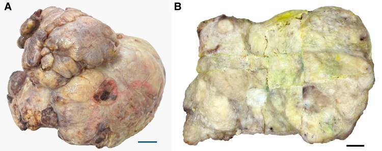
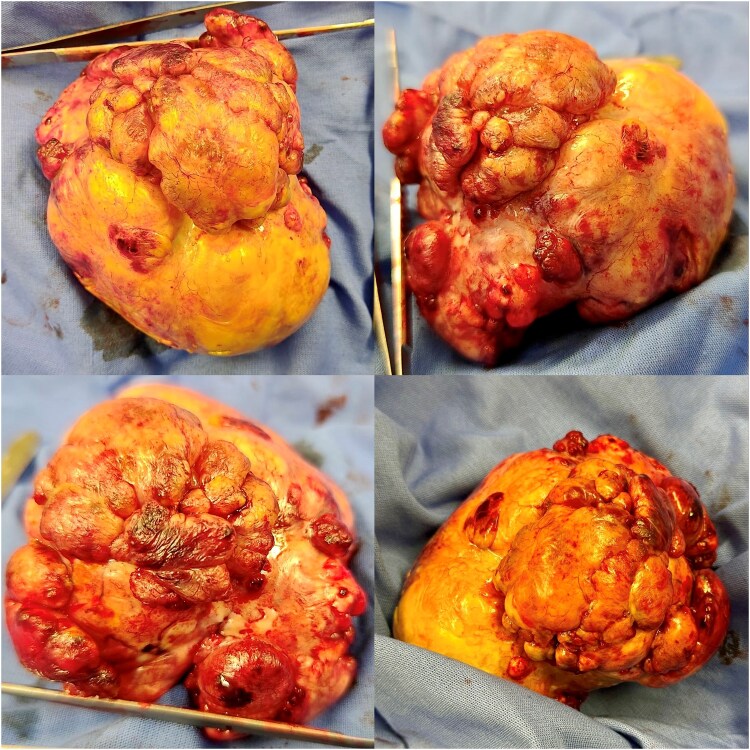
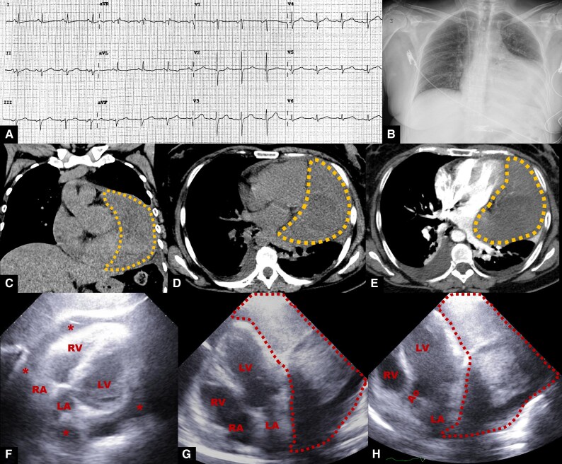
Case summary: A 56-year-old woman with a history of diabetes and active smoking presented with sudden dyspnoea. Computed tomography scan revealed a mass adjacent to the left ventricle. In addition to the large pericardial effusion, no signs of cardiac tamponade or valvulopathies were found on echocardiography. The patient underwent open-heart surgery and mass removal, finding a tumour located inside the pericardial sac and attached to the left ventricle's lateral wall; it did not invade other heart structures. Histological and immunohistochemical examination of the mass revealed SFT diagnosis. The patient was discharged from the hospital in full health, and follow-up examinations revealed no evidence of tumour recurrence.
Discussion: Solitary fibrous tumour most commonly occurs in middle-aged patients and is not gender specific. Multimodal imaging is crucial for diagnosing and managing SFT. A definitive diagnosis must be based on both immunohistochemical and histopathological findings. STAT6 immunoexpression is the most reliable marker for histopathology diagnosis. Given the high SFT recurrence rate, follow-up is essential.

 求助内容:
求助内容: 应助结果提醒方式:


