Ana Paula F C Tupynambá, Heloisa Lima Heller, Maria Fernanda L Britto, Isabella F Macedo, José A G Torres Junior, Valéria L C Borges, Ana Beatriz C Vieira, Renata Batista Ostrowski, Gustavo Henrique Soares Takano, Leonardo C G B Oliveira
{"title":"伴有眼外延伸的葡萄膜黑色素瘤进行性视力丧失1例报告。","authors":"Ana Paula F C Tupynambá, Heloisa Lima Heller, Maria Fernanda L Britto, Isabella F Macedo, José A G Torres Junior, Valéria L C Borges, Ana Beatriz C Vieira, Renata Batista Ostrowski, Gustavo Henrique Soares Takano, Leonardo C G B Oliveira","doi":"10.12659/AJCR.947979","DOIUrl":null,"url":null,"abstract":"<p><p>BACKGROUND Uveal melanoma is the most common primary intraocular malignancy in adults, often diagnosed late in resource-limited settings. The diagnosis is made through a combination of clinical ophthalmologic examination, B-mode ultrasound, and histopathological study. This report details a case of a 67-year-old woman with progressive vision loss and ocular pain due to an inferomedial uveal melanoma to highlight therapeutic limitations from delayed diagnosis. CASE REPORT A 67-year-old woman presented with 3 years of progressive vision loss, ocular pain, and a pigmented inferomedial lesion in the right eye. Examination revealed light perception vision, intraocular pressure of 38 mmHg, and a conjunctival mass with feeder vessels. Ultrasound showed a large choroidal tumor occupying 70% of the vitreous. Enucleation confirmed a 19-mm-thick mixed-cell uveal melanoma (T4a per American Joint Committee on Cancer 8th edition) with scleral invasion. Postoperative computed tomography (CT) revealed no metastases. CONCLUSIONS Delayed presentation led to enucleation as the only viable treatment. This case underscores the critical need for public awareness, early detection, and effective referral systems of pigmented ocular lesions to preserve vision, expand therapeutic options, and ensure timely access to specialized care.</p>","PeriodicalId":39064,"journal":{"name":"American Journal of Case Reports","volume":"26 ","pages":"e947979"},"PeriodicalIF":0.7000,"publicationDate":"2025-09-11","publicationTypes":"Journal Article","fieldsOfStudy":null,"isOpenAccess":false,"openAccessPdf":"https://www.ncbi.nlm.nih.gov/pmc/articles/PMC12439508/pdf/","citationCount":"0","resultStr":"{\"title\":\"Progressive Vision Loss From Uveal Melanoma with Extraocular Extension: A Case Report.\",\"authors\":\"Ana Paula F C Tupynambá, Heloisa Lima Heller, Maria Fernanda L Britto, Isabella F Macedo, José A G Torres Junior, Valéria L C Borges, Ana Beatriz C Vieira, Renata Batista Ostrowski, Gustavo Henrique Soares Takano, Leonardo C G B Oliveira\",\"doi\":\"10.12659/AJCR.947979\",\"DOIUrl\":null,\"url\":null,\"abstract\":\"<p><p>BACKGROUND Uveal melanoma is the most common primary intraocular malignancy in adults, often diagnosed late in resource-limited settings. The diagnosis is made through a combination of clinical ophthalmologic examination, B-mode ultrasound, and histopathological study. This report details a case of a 67-year-old woman with progressive vision loss and ocular pain due to an inferomedial uveal melanoma to highlight therapeutic limitations from delayed diagnosis. CASE REPORT A 67-year-old woman presented with 3 years of progressive vision loss, ocular pain, and a pigmented inferomedial lesion in the right eye. Examination revealed light perception vision, intraocular pressure of 38 mmHg, and a conjunctival mass with feeder vessels. Ultrasound showed a large choroidal tumor occupying 70% of the vitreous. Enucleation confirmed a 19-mm-thick mixed-cell uveal melanoma (T4a per American Joint Committee on Cancer 8th edition) with scleral invasion. Postoperative computed tomography (CT) revealed no metastases. CONCLUSIONS Delayed presentation led to enucleation as the only viable treatment. This case underscores the critical need for public awareness, early detection, and effective referral systems of pigmented ocular lesions to preserve vision, expand therapeutic options, and ensure timely access to specialized care.</p>\",\"PeriodicalId\":39064,\"journal\":{\"name\":\"American Journal of Case Reports\",\"volume\":\"26 \",\"pages\":\"e947979\"},\"PeriodicalIF\":0.7000,\"publicationDate\":\"2025-09-11\",\"publicationTypes\":\"Journal Article\",\"fieldsOfStudy\":null,\"isOpenAccess\":false,\"openAccessPdf\":\"https://www.ncbi.nlm.nih.gov/pmc/articles/PMC12439508/pdf/\",\"citationCount\":\"0\",\"resultStr\":null,\"platform\":\"Semanticscholar\",\"paperid\":null,\"PeriodicalName\":\"American Journal of Case Reports\",\"FirstCategoryId\":\"1085\",\"ListUrlMain\":\"https://doi.org/10.12659/AJCR.947979\",\"RegionNum\":0,\"RegionCategory\":null,\"ArticlePicture\":[],\"TitleCN\":null,\"AbstractTextCN\":null,\"PMCID\":null,\"EPubDate\":\"\",\"PubModel\":\"\",\"JCR\":\"Q3\",\"JCRName\":\"MEDICINE, GENERAL & INTERNAL\",\"Score\":null,\"Total\":0}","platform":"Semanticscholar","paperid":null,"PeriodicalName":"American Journal of Case Reports","FirstCategoryId":"1085","ListUrlMain":"https://doi.org/10.12659/AJCR.947979","RegionNum":0,"RegionCategory":null,"ArticlePicture":[],"TitleCN":null,"AbstractTextCN":null,"PMCID":null,"EPubDate":"","PubModel":"","JCR":"Q3","JCRName":"MEDICINE, GENERAL & INTERNAL","Score":null,"Total":0}
Progressive Vision Loss From Uveal Melanoma with Extraocular Extension: A Case Report.
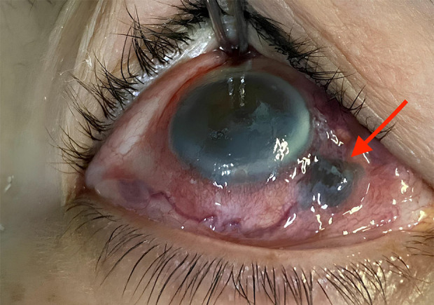
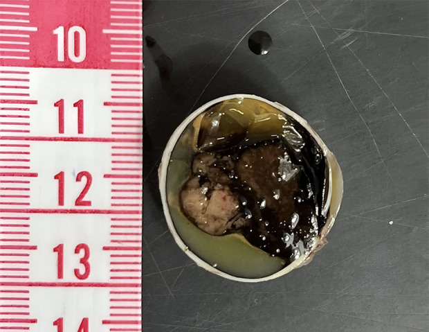
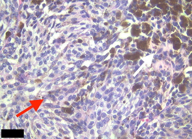
BACKGROUND Uveal melanoma is the most common primary intraocular malignancy in adults, often diagnosed late in resource-limited settings. The diagnosis is made through a combination of clinical ophthalmologic examination, B-mode ultrasound, and histopathological study. This report details a case of a 67-year-old woman with progressive vision loss and ocular pain due to an inferomedial uveal melanoma to highlight therapeutic limitations from delayed diagnosis. CASE REPORT A 67-year-old woman presented with 3 years of progressive vision loss, ocular pain, and a pigmented inferomedial lesion in the right eye. Examination revealed light perception vision, intraocular pressure of 38 mmHg, and a conjunctival mass with feeder vessels. Ultrasound showed a large choroidal tumor occupying 70% of the vitreous. Enucleation confirmed a 19-mm-thick mixed-cell uveal melanoma (T4a per American Joint Committee on Cancer 8th edition) with scleral invasion. Postoperative computed tomography (CT) revealed no metastases. CONCLUSIONS Delayed presentation led to enucleation as the only viable treatment. This case underscores the critical need for public awareness, early detection, and effective referral systems of pigmented ocular lesions to preserve vision, expand therapeutic options, and ensure timely access to specialized care.
期刊介绍:
American Journal of Case Reports is an international, peer-reviewed scientific journal that publishes single and series case reports in all medical fields. American Journal of Case Reports is issued on a continuous basis as a primary electronic journal. Print copies of a single article or a set of articles can be ordered on demand.

 求助内容:
求助内容: 应助结果提醒方式:


