Guilherme Carlos Beiruth Freire, Patricia Furtado Gonçalves, Suzana Peres Pimentel, Francisco Humberto Nociti Júnior, Márcio Zafalon Casati, Bruno César de Vasconcelos Gurgel
{"title":"裂隙缺损残余颊骨厚度对愈合部位骨整合种植体影响的体内实验研究。","authors":"Guilherme Carlos Beiruth Freire, Patricia Furtado Gonçalves, Suzana Peres Pimentel, Francisco Humberto Nociti Júnior, Márcio Zafalon Casati, Bruno César de Vasconcelos Gurgel","doi":"10.1590/1807-3107bor-2025.vol39.079","DOIUrl":null,"url":null,"abstract":"<p><p>This study aimed to histomorphometrically evaluate the effect of guided bone regeneration (GBR) and two implant surfaces on the thickness and height of newly formed bone in dehiscence defects around titanium implants. Three premolars and the first bilateral molar were extracted from ten adult mongrel dogs, and 40 buccal bone dehiscences measuring 5 mm in height and 4 mm in width were created using a University of North Carolina (UNC) periodontal probe to confirm the dimensions. Forty implants were randomly assigned to one of four groups: oxidized implant surfaces (OIS, n = 10), turned/machined implant surfaces (TIS, n = 10), OIS + GBR (n = 10), and TIS + GBR (n = 10). After 3 months, the dogs were euthanized, and the blocks containing the implants and adjacent bone were processed for non-decalcified histological analysis. Statistical analysis was performed using two-way ANOVA and the Pearson correlation (p = 0.05). The results showed that GBR significantly influenced both the vertical (height) and horizontal (thickness) dimensions of the newly formed bone (p < 0.001). Strong positive correlations were observed between the thickness and height of newly formed bone at the base of the defect, as well as between the thickness of the bone at the base of the defect and the thickness of newly formed bone in the central region of the defect. No significant correlations were found between implant surface type and bone formation. Bone regeneration occurred in both the vertical and horizontal directions, with greater bone growth in GBR-treated groups, irrespective of implant surface type (oxidized or turned).</p>","PeriodicalId":9240,"journal":{"name":"Brazilian oral research","volume":"39 ","pages":"e079"},"PeriodicalIF":1.3000,"publicationDate":"2025-09-08","publicationTypes":"Journal Article","fieldsOfStudy":null,"isOpenAccess":false,"openAccessPdf":"https://www.ncbi.nlm.nih.gov/pmc/articles/PMC12419196/pdf/","citationCount":"0","resultStr":"{\"title\":\"Influence of residual buccal bone thickness in dehiscence defects on osseointegrated dental implants in healed sites: an experimental in vivo study.\",\"authors\":\"Guilherme Carlos Beiruth Freire, Patricia Furtado Gonçalves, Suzana Peres Pimentel, Francisco Humberto Nociti Júnior, Márcio Zafalon Casati, Bruno César de Vasconcelos Gurgel\",\"doi\":\"10.1590/1807-3107bor-2025.vol39.079\",\"DOIUrl\":null,\"url\":null,\"abstract\":\"<p><p>This study aimed to histomorphometrically evaluate the effect of guided bone regeneration (GBR) and two implant surfaces on the thickness and height of newly formed bone in dehiscence defects around titanium implants. Three premolars and the first bilateral molar were extracted from ten adult mongrel dogs, and 40 buccal bone dehiscences measuring 5 mm in height and 4 mm in width were created using a University of North Carolina (UNC) periodontal probe to confirm the dimensions. Forty implants were randomly assigned to one of four groups: oxidized implant surfaces (OIS, n = 10), turned/machined implant surfaces (TIS, n = 10), OIS + GBR (n = 10), and TIS + GBR (n = 10). After 3 months, the dogs were euthanized, and the blocks containing the implants and adjacent bone were processed for non-decalcified histological analysis. Statistical analysis was performed using two-way ANOVA and the Pearson correlation (p = 0.05). The results showed that GBR significantly influenced both the vertical (height) and horizontal (thickness) dimensions of the newly formed bone (p < 0.001). Strong positive correlations were observed between the thickness and height of newly formed bone at the base of the defect, as well as between the thickness of the bone at the base of the defect and the thickness of newly formed bone in the central region of the defect. No significant correlations were found between implant surface type and bone formation. Bone regeneration occurred in both the vertical and horizontal directions, with greater bone growth in GBR-treated groups, irrespective of implant surface type (oxidized or turned).</p>\",\"PeriodicalId\":9240,\"journal\":{\"name\":\"Brazilian oral research\",\"volume\":\"39 \",\"pages\":\"e079\"},\"PeriodicalIF\":1.3000,\"publicationDate\":\"2025-09-08\",\"publicationTypes\":\"Journal Article\",\"fieldsOfStudy\":null,\"isOpenAccess\":false,\"openAccessPdf\":\"https://www.ncbi.nlm.nih.gov/pmc/articles/PMC12419196/pdf/\",\"citationCount\":\"0\",\"resultStr\":null,\"platform\":\"Semanticscholar\",\"paperid\":null,\"PeriodicalName\":\"Brazilian oral research\",\"FirstCategoryId\":\"3\",\"ListUrlMain\":\"https://doi.org/10.1590/1807-3107bor-2025.vol39.079\",\"RegionNum\":4,\"RegionCategory\":\"医学\",\"ArticlePicture\":[],\"TitleCN\":null,\"AbstractTextCN\":null,\"PMCID\":null,\"EPubDate\":\"2025/1/1 0:00:00\",\"PubModel\":\"eCollection\",\"JCR\":\"Q3\",\"JCRName\":\"DENTISTRY, ORAL SURGERY & MEDICINE\",\"Score\":null,\"Total\":0}","platform":"Semanticscholar","paperid":null,"PeriodicalName":"Brazilian oral research","FirstCategoryId":"3","ListUrlMain":"https://doi.org/10.1590/1807-3107bor-2025.vol39.079","RegionNum":4,"RegionCategory":"医学","ArticlePicture":[],"TitleCN":null,"AbstractTextCN":null,"PMCID":null,"EPubDate":"2025/1/1 0:00:00","PubModel":"eCollection","JCR":"Q3","JCRName":"DENTISTRY, ORAL SURGERY & MEDICINE","Score":null,"Total":0}
Influence of residual buccal bone thickness in dehiscence defects on osseointegrated dental implants in healed sites: an experimental in vivo study.
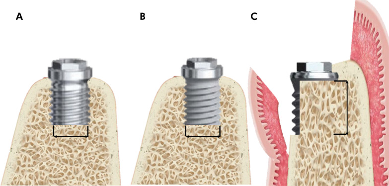
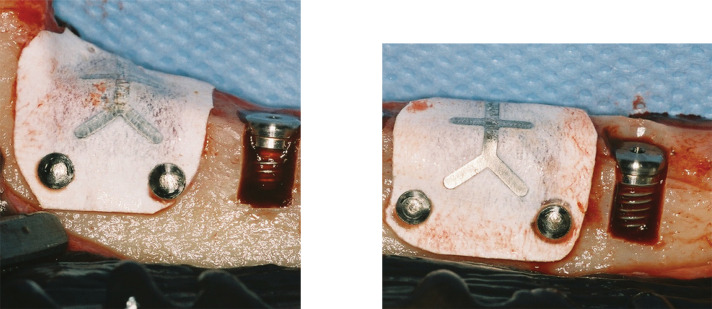
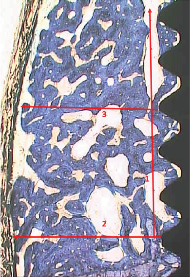
This study aimed to histomorphometrically evaluate the effect of guided bone regeneration (GBR) and two implant surfaces on the thickness and height of newly formed bone in dehiscence defects around titanium implants. Three premolars and the first bilateral molar were extracted from ten adult mongrel dogs, and 40 buccal bone dehiscences measuring 5 mm in height and 4 mm in width were created using a University of North Carolina (UNC) periodontal probe to confirm the dimensions. Forty implants were randomly assigned to one of four groups: oxidized implant surfaces (OIS, n = 10), turned/machined implant surfaces (TIS, n = 10), OIS + GBR (n = 10), and TIS + GBR (n = 10). After 3 months, the dogs were euthanized, and the blocks containing the implants and adjacent bone were processed for non-decalcified histological analysis. Statistical analysis was performed using two-way ANOVA and the Pearson correlation (p = 0.05). The results showed that GBR significantly influenced both the vertical (height) and horizontal (thickness) dimensions of the newly formed bone (p < 0.001). Strong positive correlations were observed between the thickness and height of newly formed bone at the base of the defect, as well as between the thickness of the bone at the base of the defect and the thickness of newly formed bone in the central region of the defect. No significant correlations were found between implant surface type and bone formation. Bone regeneration occurred in both the vertical and horizontal directions, with greater bone growth in GBR-treated groups, irrespective of implant surface type (oxidized or turned).
 求助内容:
求助内容: 应助结果提醒方式:


