{"title":"加强颌下腺切除:ORBEYE 3D外窥镜疗效的回顾性研究。","authors":"Masao Yagi, Mizuki Morita, Tomofumi Sakagami, Minaki Shimizu, Toshiki Utsunomiya, Kensuke Suzuki, Takuo Fujisawa, Hiroshi Iwai","doi":"10.1007/s10006-025-01446-z","DOIUrl":null,"url":null,"abstract":"<p><strong>Purpose: </strong>For submandibular gland resection, conventional surgery with the naked eye remains the standard. With its excellent automatic focus and high magnification, the ORBEYE 3D exoscope enables precise submandibular gland resection with less stress. Therefore, we aimed to examine the usefulness of the exoscope in submandibular gland resection.</p><p><strong>Methods: </strong>This retrospective study involved 12 patients who underwent submandibular gland resection with the ORBEYE exoscope at the Department of Head and Neck Surgery and Otorhinolaryngology at Kansai Medical University from April 2021 to March 2024. Surgical outcomes were retrospectively reviewed.</p><p><strong>Results: </strong>The mean age of the patients (six women and six men) was 58.5 (23-83) years. The final histopathology showed pleomorphic adenoma in eight patients, non-sebaceous-type lymphadenoma and carcinoma ex pleomorphic adenoma in one patient each, and sialolithiasis with chronic inflammation in two patients. We performed facial artery and vein sparing in all patients except one, in whom level I neck dissection, including submandibular gland excision, was performed. We identified the marginal mandibular branches of the facial, hypoglossal, and lingual nerves, as well as the submandibular duct in all patients. There were no postoperative complications in any of the patients. The mean operative time was 104 (80-129) min. The mean blood volume lost was 20 (5-43) mL.</p><p><strong>Conclusion: </strong>Using the ORBEYE exoscope in submandibular gland resection will contribute to preserving the mandibular marginal branch of the facial and lingual nerves and facial and artery veins, and visualizing the submandibular ducts, which are important structures surrounding the submandibular gland.</p>","PeriodicalId":520733,"journal":{"name":"Oral and maxillofacial surgery","volume":"29 1","pages":"149"},"PeriodicalIF":1.8000,"publicationDate":"2025-09-10","publicationTypes":"Journal Article","fieldsOfStudy":null,"isOpenAccess":false,"openAccessPdf":"https://www.ncbi.nlm.nih.gov/pmc/articles/PMC12420701/pdf/","citationCount":"0","resultStr":"{\"title\":\"Enhancing submandibular gland resection: A retrospective study on the efficacy of the ORBEYE 3D exoscope.\",\"authors\":\"Masao Yagi, Mizuki Morita, Tomofumi Sakagami, Minaki Shimizu, Toshiki Utsunomiya, Kensuke Suzuki, Takuo Fujisawa, Hiroshi Iwai\",\"doi\":\"10.1007/s10006-025-01446-z\",\"DOIUrl\":null,\"url\":null,\"abstract\":\"<p><strong>Purpose: </strong>For submandibular gland resection, conventional surgery with the naked eye remains the standard. With its excellent automatic focus and high magnification, the ORBEYE 3D exoscope enables precise submandibular gland resection with less stress. Therefore, we aimed to examine the usefulness of the exoscope in submandibular gland resection.</p><p><strong>Methods: </strong>This retrospective study involved 12 patients who underwent submandibular gland resection with the ORBEYE exoscope at the Department of Head and Neck Surgery and Otorhinolaryngology at Kansai Medical University from April 2021 to March 2024. Surgical outcomes were retrospectively reviewed.</p><p><strong>Results: </strong>The mean age of the patients (six women and six men) was 58.5 (23-83) years. The final histopathology showed pleomorphic adenoma in eight patients, non-sebaceous-type lymphadenoma and carcinoma ex pleomorphic adenoma in one patient each, and sialolithiasis with chronic inflammation in two patients. We performed facial artery and vein sparing in all patients except one, in whom level I neck dissection, including submandibular gland excision, was performed. We identified the marginal mandibular branches of the facial, hypoglossal, and lingual nerves, as well as the submandibular duct in all patients. There were no postoperative complications in any of the patients. The mean operative time was 104 (80-129) min. The mean blood volume lost was 20 (5-43) mL.</p><p><strong>Conclusion: </strong>Using the ORBEYE exoscope in submandibular gland resection will contribute to preserving the mandibular marginal branch of the facial and lingual nerves and facial and artery veins, and visualizing the submandibular ducts, which are important structures surrounding the submandibular gland.</p>\",\"PeriodicalId\":520733,\"journal\":{\"name\":\"Oral and maxillofacial surgery\",\"volume\":\"29 1\",\"pages\":\"149\"},\"PeriodicalIF\":1.8000,\"publicationDate\":\"2025-09-10\",\"publicationTypes\":\"Journal Article\",\"fieldsOfStudy\":null,\"isOpenAccess\":false,\"openAccessPdf\":\"https://www.ncbi.nlm.nih.gov/pmc/articles/PMC12420701/pdf/\",\"citationCount\":\"0\",\"resultStr\":null,\"platform\":\"Semanticscholar\",\"paperid\":null,\"PeriodicalName\":\"Oral and maxillofacial surgery\",\"FirstCategoryId\":\"1085\",\"ListUrlMain\":\"https://doi.org/10.1007/s10006-025-01446-z\",\"RegionNum\":0,\"RegionCategory\":null,\"ArticlePicture\":[],\"TitleCN\":null,\"AbstractTextCN\":null,\"PMCID\":null,\"EPubDate\":\"\",\"PubModel\":\"\",\"JCR\":\"\",\"JCRName\":\"\",\"Score\":null,\"Total\":0}","platform":"Semanticscholar","paperid":null,"PeriodicalName":"Oral and maxillofacial surgery","FirstCategoryId":"1085","ListUrlMain":"https://doi.org/10.1007/s10006-025-01446-z","RegionNum":0,"RegionCategory":null,"ArticlePicture":[],"TitleCN":null,"AbstractTextCN":null,"PMCID":null,"EPubDate":"","PubModel":"","JCR":"","JCRName":"","Score":null,"Total":0}
Enhancing submandibular gland resection: A retrospective study on the efficacy of the ORBEYE 3D exoscope.
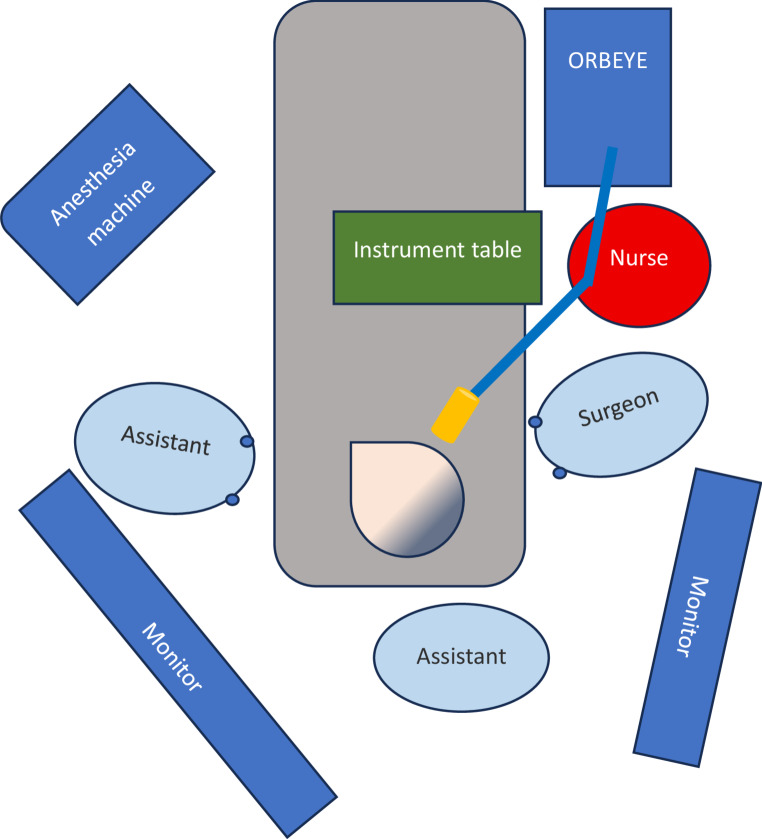
Purpose: For submandibular gland resection, conventional surgery with the naked eye remains the standard. With its excellent automatic focus and high magnification, the ORBEYE 3D exoscope enables precise submandibular gland resection with less stress. Therefore, we aimed to examine the usefulness of the exoscope in submandibular gland resection.
Methods: This retrospective study involved 12 patients who underwent submandibular gland resection with the ORBEYE exoscope at the Department of Head and Neck Surgery and Otorhinolaryngology at Kansai Medical University from April 2021 to March 2024. Surgical outcomes were retrospectively reviewed.
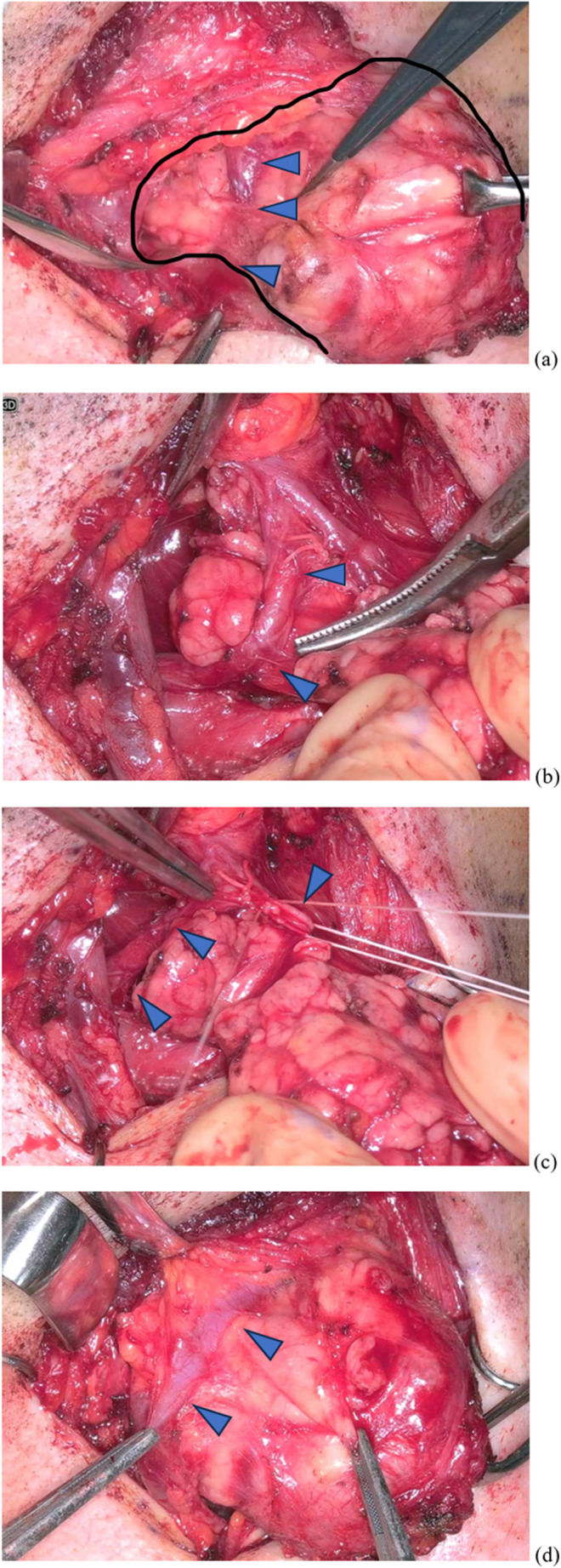
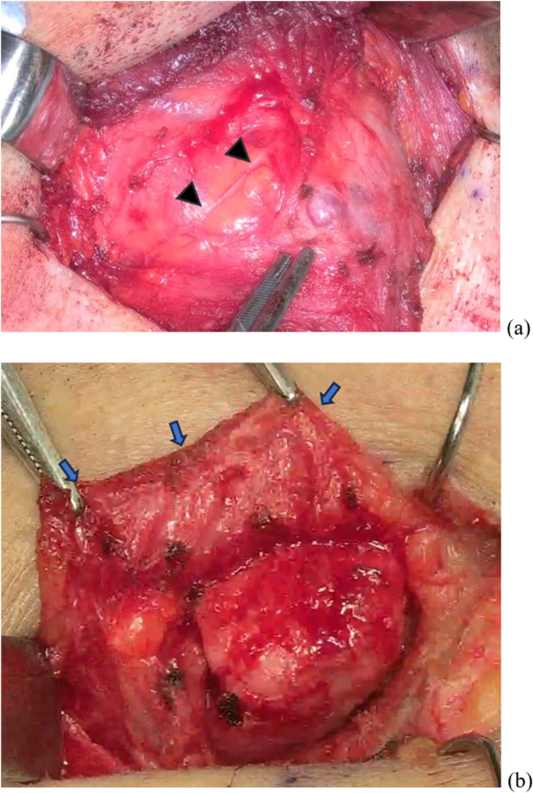
Results: The mean age of the patients (six women and six men) was 58.5 (23-83) years. The final histopathology showed pleomorphic adenoma in eight patients, non-sebaceous-type lymphadenoma and carcinoma ex pleomorphic adenoma in one patient each, and sialolithiasis with chronic inflammation in two patients. We performed facial artery and vein sparing in all patients except one, in whom level I neck dissection, including submandibular gland excision, was performed. We identified the marginal mandibular branches of the facial, hypoglossal, and lingual nerves, as well as the submandibular duct in all patients. There were no postoperative complications in any of the patients. The mean operative time was 104 (80-129) min. The mean blood volume lost was 20 (5-43) mL.
Conclusion: Using the ORBEYE exoscope in submandibular gland resection will contribute to preserving the mandibular marginal branch of the facial and lingual nerves and facial and artery veins, and visualizing the submandibular ducts, which are important structures surrounding the submandibular gland.

 求助内容:
求助内容: 应助结果提醒方式:


