{"title":"2型糖尿病黄斑血管密度与临床参数的关系","authors":"Nadia Artha Dewi, Muhammad Arfan, Herisa Rahmasari, Mutiara Kristiani Putri, Rulli Rosandi","doi":"10.2147/OPTH.S532859","DOIUrl":null,"url":null,"abstract":"<p><strong>Purpose: </strong>To evaluate macular vessel density using clinical parameters in patients with type 2 diabetes mellitus (DM) without retinopathy.</p><p><strong>Patients and methods: </strong>This cross-sectional study enrolled 32 participants (63 eyes) aged 40-60 years who met the inclusion criteria. Group 1 included 32 eyes of type 2 DM, whereas the rest had no DM. Ophthalmic examination was performed. Macular vessel density was measured using optical coherence tomography angiography (OCTA) with a 6 × 6 mm scan. Macular vessel density is correlated with patient age, HbA1c values, disease duration, and contrast sensitivity. The data were analyzed using Spearman's rank correlation and an independent <i>t</i>-test.</p><p><strong>Results: </strong>Central (foveal area) macular vessel density in the diabetic group (5.36 ± 2.87) was significantly lower than that in healthy participants (7.82 ± 3.05) (p = 0.002). The whole macular vessel density (foveal and parafoveal areas) was also lower in the DM group than in the healthy subjects (15.76 ± 3.38 vs 17.18 ± 1.92) but the difference was not statistically significant (p = 0.070). The age of DM patients has correlated with central (r = -0.522; p = 0.002) and whole macular vessel density (r = -0.369; p = 0.038). HbA1c levels, diabetes duration, and contrast sensitivity were not correlated with macular vessel density.</p><p><strong>Conclusion: </strong>Macular vessel density was lower in DM patients without retinopathy than in healthy subjects. In patients with DM, macular vessel density decreases with increasing age. We failed to find a correlation between macular vessel density and the HbA1c value, disease duration, or contrast sensitivity.</p>","PeriodicalId":93945,"journal":{"name":"Clinical ophthalmology (Auckland, N.Z.)","volume":"19 ","pages":"3113-3120"},"PeriodicalIF":0.0000,"publicationDate":"2025-09-02","publicationTypes":"Journal Article","fieldsOfStudy":null,"isOpenAccess":false,"openAccessPdf":"https://www.ncbi.nlm.nih.gov/pmc/articles/PMC12415095/pdf/","citationCount":"0","resultStr":"{\"title\":\"The Correlation Between Macular Vessel Density and Its Clinical Parameters in Diabetes Mellitus Type 2.\",\"authors\":\"Nadia Artha Dewi, Muhammad Arfan, Herisa Rahmasari, Mutiara Kristiani Putri, Rulli Rosandi\",\"doi\":\"10.2147/OPTH.S532859\",\"DOIUrl\":null,\"url\":null,\"abstract\":\"<p><strong>Purpose: </strong>To evaluate macular vessel density using clinical parameters in patients with type 2 diabetes mellitus (DM) without retinopathy.</p><p><strong>Patients and methods: </strong>This cross-sectional study enrolled 32 participants (63 eyes) aged 40-60 years who met the inclusion criteria. Group 1 included 32 eyes of type 2 DM, whereas the rest had no DM. Ophthalmic examination was performed. Macular vessel density was measured using optical coherence tomography angiography (OCTA) with a 6 × 6 mm scan. Macular vessel density is correlated with patient age, HbA1c values, disease duration, and contrast sensitivity. The data were analyzed using Spearman's rank correlation and an independent <i>t</i>-test.</p><p><strong>Results: </strong>Central (foveal area) macular vessel density in the diabetic group (5.36 ± 2.87) was significantly lower than that in healthy participants (7.82 ± 3.05) (p = 0.002). The whole macular vessel density (foveal and parafoveal areas) was also lower in the DM group than in the healthy subjects (15.76 ± 3.38 vs 17.18 ± 1.92) but the difference was not statistically significant (p = 0.070). The age of DM patients has correlated with central (r = -0.522; p = 0.002) and whole macular vessel density (r = -0.369; p = 0.038). HbA1c levels, diabetes duration, and contrast sensitivity were not correlated with macular vessel density.</p><p><strong>Conclusion: </strong>Macular vessel density was lower in DM patients without retinopathy than in healthy subjects. In patients with DM, macular vessel density decreases with increasing age. We failed to find a correlation between macular vessel density and the HbA1c value, disease duration, or contrast sensitivity.</p>\",\"PeriodicalId\":93945,\"journal\":{\"name\":\"Clinical ophthalmology (Auckland, N.Z.)\",\"volume\":\"19 \",\"pages\":\"3113-3120\"},\"PeriodicalIF\":0.0000,\"publicationDate\":\"2025-09-02\",\"publicationTypes\":\"Journal Article\",\"fieldsOfStudy\":null,\"isOpenAccess\":false,\"openAccessPdf\":\"https://www.ncbi.nlm.nih.gov/pmc/articles/PMC12415095/pdf/\",\"citationCount\":\"0\",\"resultStr\":null,\"platform\":\"Semanticscholar\",\"paperid\":null,\"PeriodicalName\":\"Clinical ophthalmology (Auckland, N.Z.)\",\"FirstCategoryId\":\"1085\",\"ListUrlMain\":\"https://doi.org/10.2147/OPTH.S532859\",\"RegionNum\":0,\"RegionCategory\":null,\"ArticlePicture\":[],\"TitleCN\":null,\"AbstractTextCN\":null,\"PMCID\":null,\"EPubDate\":\"2025/1/1 0:00:00\",\"PubModel\":\"eCollection\",\"JCR\":\"\",\"JCRName\":\"\",\"Score\":null,\"Total\":0}","platform":"Semanticscholar","paperid":null,"PeriodicalName":"Clinical ophthalmology (Auckland, N.Z.)","FirstCategoryId":"1085","ListUrlMain":"https://doi.org/10.2147/OPTH.S532859","RegionNum":0,"RegionCategory":null,"ArticlePicture":[],"TitleCN":null,"AbstractTextCN":null,"PMCID":null,"EPubDate":"2025/1/1 0:00:00","PubModel":"eCollection","JCR":"","JCRName":"","Score":null,"Total":0}
引用次数: 0
摘要
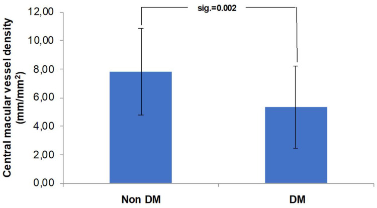
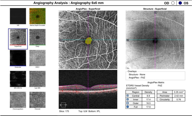
目的:应用临床参数评价无视网膜病变的2型糖尿病(DM)患者黄斑血管密度。患者和方法:本横断面研究招募了32名符合纳入标准的年龄在40-60岁的参与者(63只眼睛)。1组32只眼为2型糖尿病,其余无糖尿病。采用光学相干断层扫描血管造影(OCTA)测量6 × 6 mm扫描黄斑血管密度。黄斑血管密度与患者年龄、HbA1c值、病程和对比敏感度相关。采用Spearman秩相关和独立t检验对数据进行分析。结果:糖尿病组黄斑中央(中央凹区)血管密度(5.36±2.87)明显低于健康组(7.82±3.05)(p = 0.002)。糖尿病组黄斑血管密度(中央凹区和中央凹旁区)也低于健康组(15.76±3.38 vs 17.18±1.92),但差异无统计学意义(p = 0.070)。DM患者的年龄与黄斑中心血管密度(r = -0.522; p = 0.002)和全黄斑血管密度(r = -0.369; p = 0.038)相关。HbA1c水平、糖尿病病程和对比敏感度与黄斑血管密度无关。结论:无视网膜病变的糖尿病患者黄斑血管密度低于健康人。在糖尿病患者中,黄斑血管密度随着年龄的增长而降低。我们没有发现黄斑血管密度与HbA1c值、病程或对比敏感度之间的相关性。
The Correlation Between Macular Vessel Density and Its Clinical Parameters in Diabetes Mellitus Type 2.
Purpose: To evaluate macular vessel density using clinical parameters in patients with type 2 diabetes mellitus (DM) without retinopathy.
Patients and methods: This cross-sectional study enrolled 32 participants (63 eyes) aged 40-60 years who met the inclusion criteria. Group 1 included 32 eyes of type 2 DM, whereas the rest had no DM. Ophthalmic examination was performed. Macular vessel density was measured using optical coherence tomography angiography (OCTA) with a 6 × 6 mm scan. Macular vessel density is correlated with patient age, HbA1c values, disease duration, and contrast sensitivity. The data were analyzed using Spearman's rank correlation and an independent t-test.
Results: Central (foveal area) macular vessel density in the diabetic group (5.36 ± 2.87) was significantly lower than that in healthy participants (7.82 ± 3.05) (p = 0.002). The whole macular vessel density (foveal and parafoveal areas) was also lower in the DM group than in the healthy subjects (15.76 ± 3.38 vs 17.18 ± 1.92) but the difference was not statistically significant (p = 0.070). The age of DM patients has correlated with central (r = -0.522; p = 0.002) and whole macular vessel density (r = -0.369; p = 0.038). HbA1c levels, diabetes duration, and contrast sensitivity were not correlated with macular vessel density.
Conclusion: Macular vessel density was lower in DM patients without retinopathy than in healthy subjects. In patients with DM, macular vessel density decreases with increasing age. We failed to find a correlation between macular vessel density and the HbA1c value, disease duration, or contrast sensitivity.


 求助内容:
求助内容: 应助结果提醒方式:


