Felix L Herr, Natascha Hohmann, Christian Dascalescu, Boj Hoppe, Hannah Gildein, Verena Schäfer, Jens Ricke, Boris M Holzapfel, Lennart Schröder, Nina Hesse, Jörg Arnholdt, Paul Reidler
{"title":"3D各向同性FastView MRI定位器允许可靠的下肢扭转测量。","authors":"Felix L Herr, Natascha Hohmann, Christian Dascalescu, Boj Hoppe, Hannah Gildein, Verena Schäfer, Jens Ricke, Boris M Holzapfel, Lennart Schröder, Nina Hesse, Jörg Arnholdt, Paul Reidler","doi":"10.1186/s41747-025-00631-9","DOIUrl":null,"url":null,"abstract":"<p><p>Computed tomography (CT) and magnetic resonance imaging (MRI) are commonly used to assess femoral and tibial torsion. While CT offers high spatial resolution, it involves ionizing radiation. MRI avoids radiation but requires multiple sequences and extended acquisition time. We retrospectively evaluated whether a three-dimensional isotropic MRI localizer (FastView) could serve as a reliable and faster alternative. In this retrospective single-center study, 60 lower limbs from 30 patients, aged 27.1 ± 11.5 years (mean ± standard deviation), 19 females and 11 males, were assessed using both FastView and a dedicated MRI protocol. FastView (5 × 5 × 5 mm<sup>3</sup> voxels) imaged the entire lower limb in 17.4 s compared to nearly 7 min for the dedicated protocol. Torsion angles were measured independently by two readers. Agreement between methods was evaluated using intraclass correlation coefficients (ICCs), Bland-Altman plots, and Pearson R². No significant differences in torsion values were found (all p > 0.305). Femoral (ICC: 0.91-0.96) and tibial (ICC: 0.91-0.94) torsion showed excellent inter-modality agreement. Inter-reader reliability was also high (ICC: 0.95-0.99). Correlation values confirmed strong agreement (R²: 0.891-0.963). FastView demonstrated accuracy comparable to the dedicated protocol, offering a fast, efficient, and radiation-free option for routine torsion assessment. RELEVANCE STATEMENT: FastView MRI localizer offers a fast and resource-efficient method for assessing lower limb torsion, potentially replacing standard multisequence protocols in routine clinical practice. KEY POINTS: FastView MRI enables lower limb torsion measurements with full-limb coverage in under 20 s. Torsion angles from FastView and dedicated MRI showed no significant differences. Femoral and tibial ICCs between 0.91 and 0.96 confirm excellent inter-protocol agreement. Inter-reader agreement was consistently high across both protocols. FastView may replace multisequence MRI protocols in routine clinical torsion assessment.</p>","PeriodicalId":36926,"journal":{"name":"European Radiology Experimental","volume":"9 1","pages":"87"},"PeriodicalIF":3.6000,"publicationDate":"2025-09-09","publicationTypes":"Journal Article","fieldsOfStudy":null,"isOpenAccess":false,"openAccessPdf":"https://www.ncbi.nlm.nih.gov/pmc/articles/PMC12420559/pdf/","citationCount":"0","resultStr":"{\"title\":\"3D isotropic FastView MRI localizer allows reliable torsion measurements of the lower limb.\",\"authors\":\"Felix L Herr, Natascha Hohmann, Christian Dascalescu, Boj Hoppe, Hannah Gildein, Verena Schäfer, Jens Ricke, Boris M Holzapfel, Lennart Schröder, Nina Hesse, Jörg Arnholdt, Paul Reidler\",\"doi\":\"10.1186/s41747-025-00631-9\",\"DOIUrl\":null,\"url\":null,\"abstract\":\"<p><p>Computed tomography (CT) and magnetic resonance imaging (MRI) are commonly used to assess femoral and tibial torsion. While CT offers high spatial resolution, it involves ionizing radiation. MRI avoids radiation but requires multiple sequences and extended acquisition time. We retrospectively evaluated whether a three-dimensional isotropic MRI localizer (FastView) could serve as a reliable and faster alternative. In this retrospective single-center study, 60 lower limbs from 30 patients, aged 27.1 ± 11.5 years (mean ± standard deviation), 19 females and 11 males, were assessed using both FastView and a dedicated MRI protocol. FastView (5 × 5 × 5 mm<sup>3</sup> voxels) imaged the entire lower limb in 17.4 s compared to nearly 7 min for the dedicated protocol. Torsion angles were measured independently by two readers. Agreement between methods was evaluated using intraclass correlation coefficients (ICCs), Bland-Altman plots, and Pearson R². No significant differences in torsion values were found (all p > 0.305). Femoral (ICC: 0.91-0.96) and tibial (ICC: 0.91-0.94) torsion showed excellent inter-modality agreement. Inter-reader reliability was also high (ICC: 0.95-0.99). Correlation values confirmed strong agreement (R²: 0.891-0.963). FastView demonstrated accuracy comparable to the dedicated protocol, offering a fast, efficient, and radiation-free option for routine torsion assessment. RELEVANCE STATEMENT: FastView MRI localizer offers a fast and resource-efficient method for assessing lower limb torsion, potentially replacing standard multisequence protocols in routine clinical practice. KEY POINTS: FastView MRI enables lower limb torsion measurements with full-limb coverage in under 20 s. Torsion angles from FastView and dedicated MRI showed no significant differences. Femoral and tibial ICCs between 0.91 and 0.96 confirm excellent inter-protocol agreement. Inter-reader agreement was consistently high across both protocols. FastView may replace multisequence MRI protocols in routine clinical torsion assessment.</p>\",\"PeriodicalId\":36926,\"journal\":{\"name\":\"European Radiology Experimental\",\"volume\":\"9 1\",\"pages\":\"87\"},\"PeriodicalIF\":3.6000,\"publicationDate\":\"2025-09-09\",\"publicationTypes\":\"Journal Article\",\"fieldsOfStudy\":null,\"isOpenAccess\":false,\"openAccessPdf\":\"https://www.ncbi.nlm.nih.gov/pmc/articles/PMC12420559/pdf/\",\"citationCount\":\"0\",\"resultStr\":null,\"platform\":\"Semanticscholar\",\"paperid\":null,\"PeriodicalName\":\"European Radiology Experimental\",\"FirstCategoryId\":\"1085\",\"ListUrlMain\":\"https://doi.org/10.1186/s41747-025-00631-9\",\"RegionNum\":0,\"RegionCategory\":null,\"ArticlePicture\":[],\"TitleCN\":null,\"AbstractTextCN\":null,\"PMCID\":null,\"EPubDate\":\"\",\"PubModel\":\"\",\"JCR\":\"Q1\",\"JCRName\":\"RADIOLOGY, NUCLEAR MEDICINE & MEDICAL IMAGING\",\"Score\":null,\"Total\":0}","platform":"Semanticscholar","paperid":null,"PeriodicalName":"European Radiology Experimental","FirstCategoryId":"1085","ListUrlMain":"https://doi.org/10.1186/s41747-025-00631-9","RegionNum":0,"RegionCategory":null,"ArticlePicture":[],"TitleCN":null,"AbstractTextCN":null,"PMCID":null,"EPubDate":"","PubModel":"","JCR":"Q1","JCRName":"RADIOLOGY, NUCLEAR MEDICINE & MEDICAL IMAGING","Score":null,"Total":0}
3D isotropic FastView MRI localizer allows reliable torsion measurements of the lower limb.
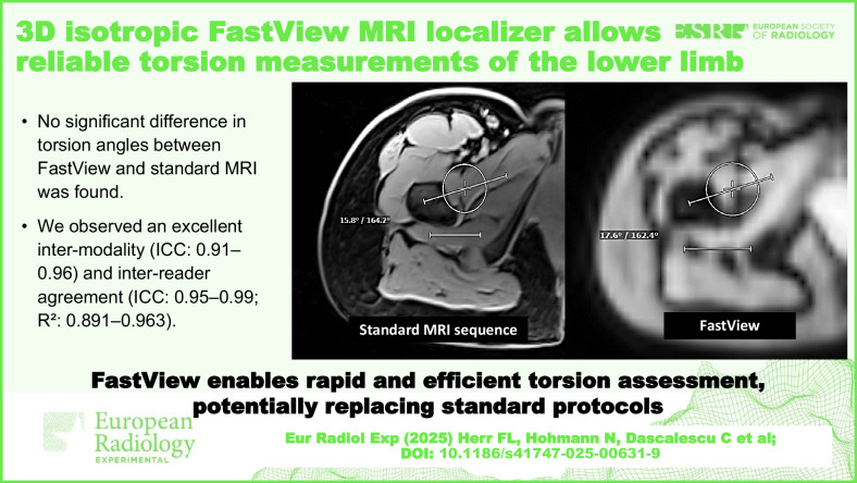
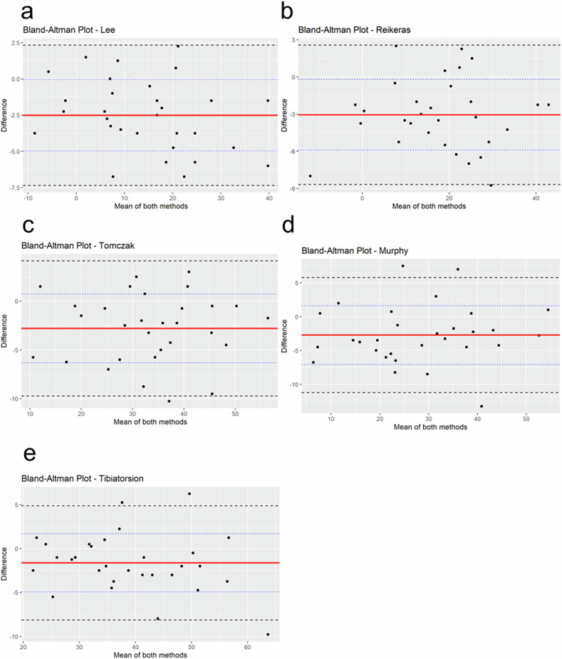
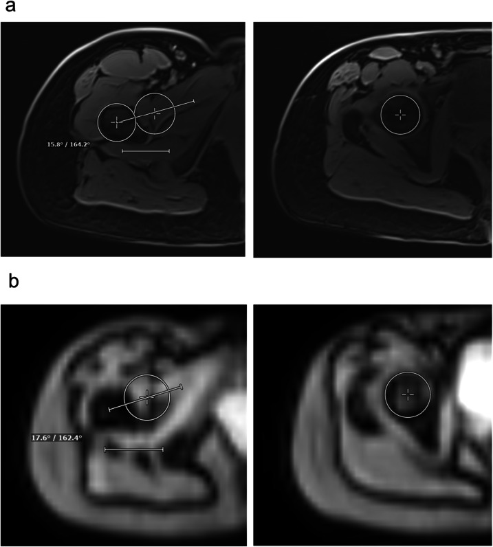
Computed tomography (CT) and magnetic resonance imaging (MRI) are commonly used to assess femoral and tibial torsion. While CT offers high spatial resolution, it involves ionizing radiation. MRI avoids radiation but requires multiple sequences and extended acquisition time. We retrospectively evaluated whether a three-dimensional isotropic MRI localizer (FastView) could serve as a reliable and faster alternative. In this retrospective single-center study, 60 lower limbs from 30 patients, aged 27.1 ± 11.5 years (mean ± standard deviation), 19 females and 11 males, were assessed using both FastView and a dedicated MRI protocol. FastView (5 × 5 × 5 mm3 voxels) imaged the entire lower limb in 17.4 s compared to nearly 7 min for the dedicated protocol. Torsion angles were measured independently by two readers. Agreement between methods was evaluated using intraclass correlation coefficients (ICCs), Bland-Altman plots, and Pearson R². No significant differences in torsion values were found (all p > 0.305). Femoral (ICC: 0.91-0.96) and tibial (ICC: 0.91-0.94) torsion showed excellent inter-modality agreement. Inter-reader reliability was also high (ICC: 0.95-0.99). Correlation values confirmed strong agreement (R²: 0.891-0.963). FastView demonstrated accuracy comparable to the dedicated protocol, offering a fast, efficient, and radiation-free option for routine torsion assessment. RELEVANCE STATEMENT: FastView MRI localizer offers a fast and resource-efficient method for assessing lower limb torsion, potentially replacing standard multisequence protocols in routine clinical practice. KEY POINTS: FastView MRI enables lower limb torsion measurements with full-limb coverage in under 20 s. Torsion angles from FastView and dedicated MRI showed no significant differences. Femoral and tibial ICCs between 0.91 and 0.96 confirm excellent inter-protocol agreement. Inter-reader agreement was consistently high across both protocols. FastView may replace multisequence MRI protocols in routine clinical torsion assessment.

 求助内容:
求助内容: 应助结果提醒方式:


