Nor Hasniza Abdul Halim, Idris Ibrahim, Nadia Mohamed Tarmizi, Mohd Hazeman Zakaria
{"title":"骨显像和单光子发射计算机断层扫描(SPECT)显示肋骨纤维发育不良的香蕉样外观。","authors":"Nor Hasniza Abdul Halim, Idris Ibrahim, Nadia Mohamed Tarmizi, Mohd Hazeman Zakaria","doi":"10.4103/ijnm.ijnm_141_24","DOIUrl":null,"url":null,"abstract":"<p><p>The patient, a 37-year-old male, initially presented with per rectal bleed. Colonoscopy revealed a circumferential lesion within the lower rectum, along with a few satellite lesions. At that time, we performed a biopsy, but the histopathological examination revealed consistent solitary rectal ulcer syndrome. Subsequent bone scan revealed increased radiotracer uptake at the left 6<sup>th</sup> rib, resulting in a \"banana-like appearance\" and a ground glass appearance on the corresponding single-photon emission computed tomography (SPECT), in keeping with fibrous dysplasia. Given his asymptomatic state, the patient received reassurance and surveillance regarding his benign bone lesion.</p>","PeriodicalId":45830,"journal":{"name":"Indian Journal of Nuclear Medicine","volume":"40 3","pages":"172-173"},"PeriodicalIF":0.5000,"publicationDate":"2025-05-01","publicationTypes":"Journal Article","fieldsOfStudy":null,"isOpenAccess":false,"openAccessPdf":"https://www.ncbi.nlm.nih.gov/pmc/articles/PMC12416581/pdf/","citationCount":"0","resultStr":"{\"title\":\"Banana-like Appearance of Rib Fibrous Dysplasia in Bone Scintigraphy and Single-Photon Emission Computed Tomography (SPECT).\",\"authors\":\"Nor Hasniza Abdul Halim, Idris Ibrahim, Nadia Mohamed Tarmizi, Mohd Hazeman Zakaria\",\"doi\":\"10.4103/ijnm.ijnm_141_24\",\"DOIUrl\":null,\"url\":null,\"abstract\":\"<p><p>The patient, a 37-year-old male, initially presented with per rectal bleed. Colonoscopy revealed a circumferential lesion within the lower rectum, along with a few satellite lesions. At that time, we performed a biopsy, but the histopathological examination revealed consistent solitary rectal ulcer syndrome. Subsequent bone scan revealed increased radiotracer uptake at the left 6<sup>th</sup> rib, resulting in a \\\"banana-like appearance\\\" and a ground glass appearance on the corresponding single-photon emission computed tomography (SPECT), in keeping with fibrous dysplasia. Given his asymptomatic state, the patient received reassurance and surveillance regarding his benign bone lesion.</p>\",\"PeriodicalId\":45830,\"journal\":{\"name\":\"Indian Journal of Nuclear Medicine\",\"volume\":\"40 3\",\"pages\":\"172-173\"},\"PeriodicalIF\":0.5000,\"publicationDate\":\"2025-05-01\",\"publicationTypes\":\"Journal Article\",\"fieldsOfStudy\":null,\"isOpenAccess\":false,\"openAccessPdf\":\"https://www.ncbi.nlm.nih.gov/pmc/articles/PMC12416581/pdf/\",\"citationCount\":\"0\",\"resultStr\":null,\"platform\":\"Semanticscholar\",\"paperid\":null,\"PeriodicalName\":\"Indian Journal of Nuclear Medicine\",\"FirstCategoryId\":\"1085\",\"ListUrlMain\":\"https://doi.org/10.4103/ijnm.ijnm_141_24\",\"RegionNum\":0,\"RegionCategory\":null,\"ArticlePicture\":[],\"TitleCN\":null,\"AbstractTextCN\":null,\"PMCID\":null,\"EPubDate\":\"2025/8/7 0:00:00\",\"PubModel\":\"Epub\",\"JCR\":\"Q4\",\"JCRName\":\"RADIOLOGY, NUCLEAR MEDICINE & MEDICAL IMAGING\",\"Score\":null,\"Total\":0}","platform":"Semanticscholar","paperid":null,"PeriodicalName":"Indian Journal of Nuclear Medicine","FirstCategoryId":"1085","ListUrlMain":"https://doi.org/10.4103/ijnm.ijnm_141_24","RegionNum":0,"RegionCategory":null,"ArticlePicture":[],"TitleCN":null,"AbstractTextCN":null,"PMCID":null,"EPubDate":"2025/8/7 0:00:00","PubModel":"Epub","JCR":"Q4","JCRName":"RADIOLOGY, NUCLEAR MEDICINE & MEDICAL IMAGING","Score":null,"Total":0}
Banana-like Appearance of Rib Fibrous Dysplasia in Bone Scintigraphy and Single-Photon Emission Computed Tomography (SPECT).
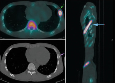
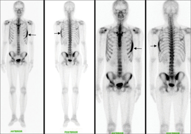
The patient, a 37-year-old male, initially presented with per rectal bleed. Colonoscopy revealed a circumferential lesion within the lower rectum, along with a few satellite lesions. At that time, we performed a biopsy, but the histopathological examination revealed consistent solitary rectal ulcer syndrome. Subsequent bone scan revealed increased radiotracer uptake at the left 6th rib, resulting in a "banana-like appearance" and a ground glass appearance on the corresponding single-photon emission computed tomography (SPECT), in keeping with fibrous dysplasia. Given his asymptomatic state, the patient received reassurance and surveillance regarding his benign bone lesion.

 求助内容:
求助内容: 应助结果提醒方式:


