Sambit Sagar, Dikhra Khan, Rahul Viswanathan, Prateek Kaushik, Rakesh Kumar
{"title":"FDG PET-CT显示罕见的面部血管肉瘤转移到肝脏和脾脏。","authors":"Sambit Sagar, Dikhra Khan, Rahul Viswanathan, Prateek Kaushik, Rakesh Kumar","doi":"10.4103/ijnm.ijnm_104_22","DOIUrl":null,"url":null,"abstract":"<p><p>Angiosarcoma is a rare type of soft-tissue sarcoma, constituting only 1% out of all soft-tissue sarcomas pathologically originating from lymphatic or vascular endothelial cells. Angiosarcomas are reported to be very aggressive with a high incidence of metastases to different sites; therefore, it is very important to determine disease extension and detect local recurrence and/or distant metastases for appropriate management. We report a case of a 55-year-old Indian male who presented with soft-tissue thickening of the left cheek for which biopsy revealed angiosarcoma and was referred for fludeoxyglucose positron emission tomography/computed tomography (FDG PET/CT) to assess the extent of disease highlighting the potential role of FDG PET/CT in rare malignancies like angiosarcomas.</p>","PeriodicalId":45830,"journal":{"name":"Indian Journal of Nuclear Medicine","volume":"40 3","pages":"168-169"},"PeriodicalIF":0.5000,"publicationDate":"2025-05-01","publicationTypes":"Journal Article","fieldsOfStudy":null,"isOpenAccess":false,"openAccessPdf":"https://www.ncbi.nlm.nih.gov/pmc/articles/PMC12416574/pdf/","citationCount":"0","resultStr":"{\"title\":\"Rare Metastases to Liver and Spleen form a Seldom Case of Facial Angiosarcoma Demonstrated on Staging FDG PET-CT.\",\"authors\":\"Sambit Sagar, Dikhra Khan, Rahul Viswanathan, Prateek Kaushik, Rakesh Kumar\",\"doi\":\"10.4103/ijnm.ijnm_104_22\",\"DOIUrl\":null,\"url\":null,\"abstract\":\"<p><p>Angiosarcoma is a rare type of soft-tissue sarcoma, constituting only 1% out of all soft-tissue sarcomas pathologically originating from lymphatic or vascular endothelial cells. Angiosarcomas are reported to be very aggressive with a high incidence of metastases to different sites; therefore, it is very important to determine disease extension and detect local recurrence and/or distant metastases for appropriate management. We report a case of a 55-year-old Indian male who presented with soft-tissue thickening of the left cheek for which biopsy revealed angiosarcoma and was referred for fludeoxyglucose positron emission tomography/computed tomography (FDG PET/CT) to assess the extent of disease highlighting the potential role of FDG PET/CT in rare malignancies like angiosarcomas.</p>\",\"PeriodicalId\":45830,\"journal\":{\"name\":\"Indian Journal of Nuclear Medicine\",\"volume\":\"40 3\",\"pages\":\"168-169\"},\"PeriodicalIF\":0.5000,\"publicationDate\":\"2025-05-01\",\"publicationTypes\":\"Journal Article\",\"fieldsOfStudy\":null,\"isOpenAccess\":false,\"openAccessPdf\":\"https://www.ncbi.nlm.nih.gov/pmc/articles/PMC12416574/pdf/\",\"citationCount\":\"0\",\"resultStr\":null,\"platform\":\"Semanticscholar\",\"paperid\":null,\"PeriodicalName\":\"Indian Journal of Nuclear Medicine\",\"FirstCategoryId\":\"1085\",\"ListUrlMain\":\"https://doi.org/10.4103/ijnm.ijnm_104_22\",\"RegionNum\":0,\"RegionCategory\":null,\"ArticlePicture\":[],\"TitleCN\":null,\"AbstractTextCN\":null,\"PMCID\":null,\"EPubDate\":\"2025/8/7 0:00:00\",\"PubModel\":\"Epub\",\"JCR\":\"Q4\",\"JCRName\":\"RADIOLOGY, NUCLEAR MEDICINE & MEDICAL IMAGING\",\"Score\":null,\"Total\":0}","platform":"Semanticscholar","paperid":null,"PeriodicalName":"Indian Journal of Nuclear Medicine","FirstCategoryId":"1085","ListUrlMain":"https://doi.org/10.4103/ijnm.ijnm_104_22","RegionNum":0,"RegionCategory":null,"ArticlePicture":[],"TitleCN":null,"AbstractTextCN":null,"PMCID":null,"EPubDate":"2025/8/7 0:00:00","PubModel":"Epub","JCR":"Q4","JCRName":"RADIOLOGY, NUCLEAR MEDICINE & MEDICAL IMAGING","Score":null,"Total":0}
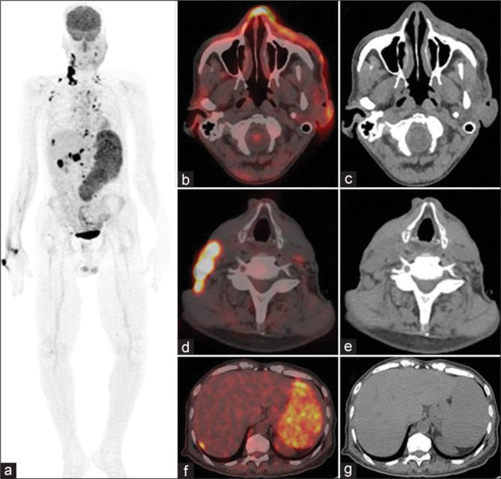
Rare Metastases to Liver and Spleen form a Seldom Case of Facial Angiosarcoma Demonstrated on Staging FDG PET-CT.
Angiosarcoma is a rare type of soft-tissue sarcoma, constituting only 1% out of all soft-tissue sarcomas pathologically originating from lymphatic or vascular endothelial cells. Angiosarcomas are reported to be very aggressive with a high incidence of metastases to different sites; therefore, it is very important to determine disease extension and detect local recurrence and/or distant metastases for appropriate management. We report a case of a 55-year-old Indian male who presented with soft-tissue thickening of the left cheek for which biopsy revealed angiosarcoma and was referred for fludeoxyglucose positron emission tomography/computed tomography (FDG PET/CT) to assess the extent of disease highlighting the potential role of FDG PET/CT in rare malignancies like angiosarcomas.

 求助内容:
求助内容: 应助结果提醒方式:


