John Pathak, Dikhra Khan, Shamim Ahmed Shamim, Sambit Sagar, Sameer Rastogi
{"title":"探讨FDG PET-CT在转移性复发骨外肾骨肉瘤中的应用。","authors":"John Pathak, Dikhra Khan, Shamim Ahmed Shamim, Sambit Sagar, Sameer Rastogi","doi":"10.4103/ijnm.ijnm_83_22","DOIUrl":null,"url":null,"abstract":"<p><p>Metastatic renal osteosarcoma is a rare entity. We report a case of a 52-year-old male postright nephrectomy status presented to us with metastatic renal osteosarcoma. 18-fluorine- fluorodeoxyglucose (<sup>18</sup>F-FDG) avid lesions were seen in the right renal bed with extension to adjacent hepatic parenchyma. The patient exhibited a favorable response to 6 cycles of carboplatin and doxorubicin with no FDG avid lesions in the postchemotherapy follow-up scan. On 6-month follow-up scan, scan findings reveal a recurrent lesion at the renal bed region infiltrating psoas muscle and liver metastatic lesion. This case highlights the use of <sup>18</sup>F-FDG positron emission tomography/computed tomography in metastatic recurrent extraskeletal osteosarcoma.</p>","PeriodicalId":45830,"journal":{"name":"Indian Journal of Nuclear Medicine","volume":"40 3","pages":"166-167"},"PeriodicalIF":0.5000,"publicationDate":"2025-05-01","publicationTypes":"Journal Article","fieldsOfStudy":null,"isOpenAccess":false,"openAccessPdf":"https://www.ncbi.nlm.nih.gov/pmc/articles/PMC12416627/pdf/","citationCount":"0","resultStr":"{\"title\":\"Exploring the Utility of FDG PET-CT in Metastatic Recurrent Extra Skeletal Renal Osteosarcoma.\",\"authors\":\"John Pathak, Dikhra Khan, Shamim Ahmed Shamim, Sambit Sagar, Sameer Rastogi\",\"doi\":\"10.4103/ijnm.ijnm_83_22\",\"DOIUrl\":null,\"url\":null,\"abstract\":\"<p><p>Metastatic renal osteosarcoma is a rare entity. We report a case of a 52-year-old male postright nephrectomy status presented to us with metastatic renal osteosarcoma. 18-fluorine- fluorodeoxyglucose (<sup>18</sup>F-FDG) avid lesions were seen in the right renal bed with extension to adjacent hepatic parenchyma. The patient exhibited a favorable response to 6 cycles of carboplatin and doxorubicin with no FDG avid lesions in the postchemotherapy follow-up scan. On 6-month follow-up scan, scan findings reveal a recurrent lesion at the renal bed region infiltrating psoas muscle and liver metastatic lesion. This case highlights the use of <sup>18</sup>F-FDG positron emission tomography/computed tomography in metastatic recurrent extraskeletal osteosarcoma.</p>\",\"PeriodicalId\":45830,\"journal\":{\"name\":\"Indian Journal of Nuclear Medicine\",\"volume\":\"40 3\",\"pages\":\"166-167\"},\"PeriodicalIF\":0.5000,\"publicationDate\":\"2025-05-01\",\"publicationTypes\":\"Journal Article\",\"fieldsOfStudy\":null,\"isOpenAccess\":false,\"openAccessPdf\":\"https://www.ncbi.nlm.nih.gov/pmc/articles/PMC12416627/pdf/\",\"citationCount\":\"0\",\"resultStr\":null,\"platform\":\"Semanticscholar\",\"paperid\":null,\"PeriodicalName\":\"Indian Journal of Nuclear Medicine\",\"FirstCategoryId\":\"1085\",\"ListUrlMain\":\"https://doi.org/10.4103/ijnm.ijnm_83_22\",\"RegionNum\":0,\"RegionCategory\":null,\"ArticlePicture\":[],\"TitleCN\":null,\"AbstractTextCN\":null,\"PMCID\":null,\"EPubDate\":\"2025/8/7 0:00:00\",\"PubModel\":\"Epub\",\"JCR\":\"Q4\",\"JCRName\":\"RADIOLOGY, NUCLEAR MEDICINE & MEDICAL IMAGING\",\"Score\":null,\"Total\":0}","platform":"Semanticscholar","paperid":null,"PeriodicalName":"Indian Journal of Nuclear Medicine","FirstCategoryId":"1085","ListUrlMain":"https://doi.org/10.4103/ijnm.ijnm_83_22","RegionNum":0,"RegionCategory":null,"ArticlePicture":[],"TitleCN":null,"AbstractTextCN":null,"PMCID":null,"EPubDate":"2025/8/7 0:00:00","PubModel":"Epub","JCR":"Q4","JCRName":"RADIOLOGY, NUCLEAR MEDICINE & MEDICAL IMAGING","Score":null,"Total":0}
Exploring the Utility of FDG PET-CT in Metastatic Recurrent Extra Skeletal Renal Osteosarcoma.
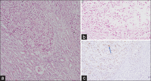
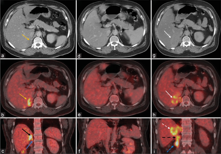
Metastatic renal osteosarcoma is a rare entity. We report a case of a 52-year-old male postright nephrectomy status presented to us with metastatic renal osteosarcoma. 18-fluorine- fluorodeoxyglucose (18F-FDG) avid lesions were seen in the right renal bed with extension to adjacent hepatic parenchyma. The patient exhibited a favorable response to 6 cycles of carboplatin and doxorubicin with no FDG avid lesions in the postchemotherapy follow-up scan. On 6-month follow-up scan, scan findings reveal a recurrent lesion at the renal bed region infiltrating psoas muscle and liver metastatic lesion. This case highlights the use of 18F-FDG positron emission tomography/computed tomography in metastatic recurrent extraskeletal osteosarcoma.

 求助内容:
求助内容: 应助结果提醒方式:


