Tc-99m MDP在多灶非典型骨化纤维黏液样瘤中的活性升高。

IF 0.5 Q4 RADIOLOGY, NUCLEAR MEDICINE & MEDICAL IMAGING
Indian Journal of Nuclear Medicine Pub Date : 2025-05-01 Epub Date: 2025-08-07 DOI:10.4103/ijnm.ijnm_28_25
Hui Ming, Yuanhao Chen, Xun Sun, Hui Yu
{"title":"Tc-99m MDP在多灶非典型骨化纤维黏液样瘤中的活性升高。","authors":"Hui Ming, Yuanhao Chen, Xun Sun, Hui Yu","doi":"10.4103/ijnm.ijnm_28_25","DOIUrl":null,"url":null,"abstract":"<p><p>This case demonstrates extraosseous technetium-99m methylene diphosphonate (Tc-99m MDP) accumulation from an atypical ossifying fibromyxoid tumor (OFMT). A 63-year-old man presented with a 10-year history of a gradually enlarging, painless back mass. Physical examination revealed multiple hard, nontender subcutaneous nodules without signs of inflammation or pigmentation. Computed tomography scan of the back showed calcified masses. Whole-body bone scintigraphy revealed areas of increased Tc-99m MDP uptake in subcutaneous regions, suggesting extraosseous uptake. Histopathological examination of the back mass confirmed the diagnosis of atypical OFMT.</p>","PeriodicalId":45830,"journal":{"name":"Indian Journal of Nuclear Medicine","volume":"40 3","pages":"181-182"},"PeriodicalIF":0.5000,"publicationDate":"2025-05-01","publicationTypes":"Journal Article","fieldsOfStudy":null,"isOpenAccess":false,"openAccessPdf":"https://www.ncbi.nlm.nih.gov/pmc/articles/PMC12416612/pdf/","citationCount":"0","resultStr":"{\"title\":\"Increased Tc-99m MDP Activities in Multifocal Atypical Ossifying Fibromyxoid Tumor.\",\"authors\":\"Hui Ming, Yuanhao Chen, Xun Sun, Hui Yu\",\"doi\":\"10.4103/ijnm.ijnm_28_25\",\"DOIUrl\":null,\"url\":null,\"abstract\":\"<p><p>This case demonstrates extraosseous technetium-99m methylene diphosphonate (Tc-99m MDP) accumulation from an atypical ossifying fibromyxoid tumor (OFMT). A 63-year-old man presented with a 10-year history of a gradually enlarging, painless back mass. Physical examination revealed multiple hard, nontender subcutaneous nodules without signs of inflammation or pigmentation. Computed tomography scan of the back showed calcified masses. Whole-body bone scintigraphy revealed areas of increased Tc-99m MDP uptake in subcutaneous regions, suggesting extraosseous uptake. Histopathological examination of the back mass confirmed the diagnosis of atypical OFMT.</p>\",\"PeriodicalId\":45830,\"journal\":{\"name\":\"Indian Journal of Nuclear Medicine\",\"volume\":\"40 3\",\"pages\":\"181-182\"},\"PeriodicalIF\":0.5000,\"publicationDate\":\"2025-05-01\",\"publicationTypes\":\"Journal Article\",\"fieldsOfStudy\":null,\"isOpenAccess\":false,\"openAccessPdf\":\"https://www.ncbi.nlm.nih.gov/pmc/articles/PMC12416612/pdf/\",\"citationCount\":\"0\",\"resultStr\":null,\"platform\":\"Semanticscholar\",\"paperid\":null,\"PeriodicalName\":\"Indian Journal of Nuclear Medicine\",\"FirstCategoryId\":\"1085\",\"ListUrlMain\":\"https://doi.org/10.4103/ijnm.ijnm_28_25\",\"RegionNum\":0,\"RegionCategory\":null,\"ArticlePicture\":[],\"TitleCN\":null,\"AbstractTextCN\":null,\"PMCID\":null,\"EPubDate\":\"2025/8/7 0:00:00\",\"PubModel\":\"Epub\",\"JCR\":\"Q4\",\"JCRName\":\"RADIOLOGY, NUCLEAR MEDICINE & MEDICAL IMAGING\",\"Score\":null,\"Total\":0}","platform":"Semanticscholar","paperid":null,"PeriodicalName":"Indian Journal of Nuclear Medicine","FirstCategoryId":"1085","ListUrlMain":"https://doi.org/10.4103/ijnm.ijnm_28_25","RegionNum":0,"RegionCategory":null,"ArticlePicture":[],"TitleCN":null,"AbstractTextCN":null,"PMCID":null,"EPubDate":"2025/8/7 0:00:00","PubModel":"Epub","JCR":"Q4","JCRName":"RADIOLOGY, NUCLEAR MEDICINE & MEDICAL IMAGING","Score":null,"Total":0}
引用次数: 0

摘要

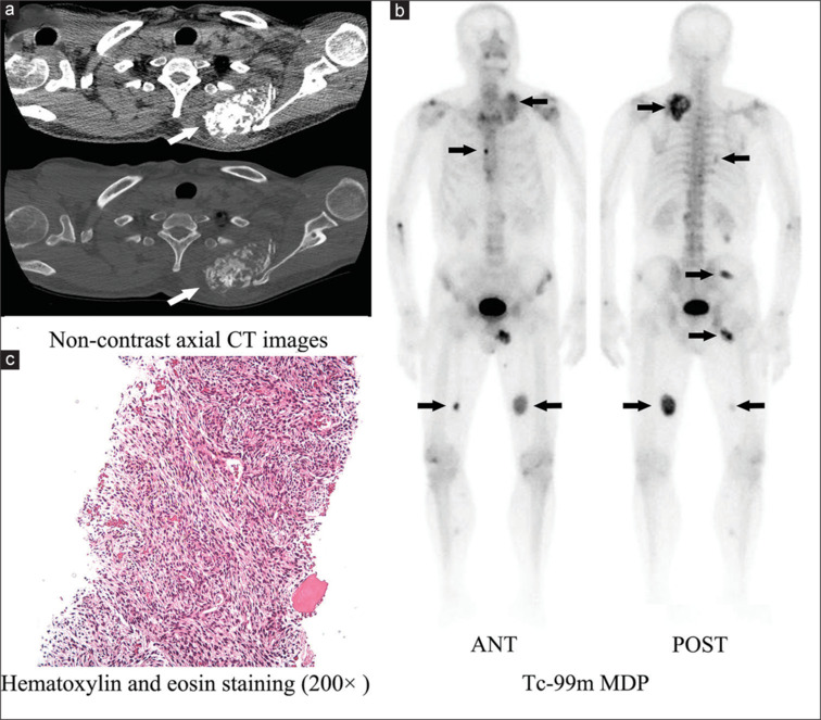
本病例显示非典型骨化纤维黏液样肿瘤(OFMT)的骨外锝-99m二膦酸亚甲基(Tc-99m MDP)积累。男性,63岁,有10年逐渐增大的无痛性背部肿块病史。体格检查显示多发坚硬、无压痛的皮下结节,无炎症或色素沉着迹象。背部计算机断层扫描显示钙化肿块。全身骨显像显示皮下区域Tc-99m MDP摄取增加,提示骨外摄取。背部肿块的组织病理学检查证实了非典型OFMT的诊断。
本文章由计算机程序翻译,如有差异,请以英文原文为准。

Increased Tc-99m MDP Activities in Multifocal Atypical Ossifying Fibromyxoid Tumor.

Increased Tc-99m MDP Activities in Multifocal Atypical Ossifying Fibromyxoid Tumor.

This case demonstrates extraosseous technetium-99m methylene diphosphonate (Tc-99m MDP) accumulation from an atypical ossifying fibromyxoid tumor (OFMT). A 63-year-old man presented with a 10-year history of a gradually enlarging, painless back mass. Physical examination revealed multiple hard, nontender subcutaneous nodules without signs of inflammation or pigmentation. Computed tomography scan of the back showed calcified masses. Whole-body bone scintigraphy revealed areas of increased Tc-99m MDP uptake in subcutaneous regions, suggesting extraosseous uptake. Histopathological examination of the back mass confirmed the diagnosis of atypical OFMT.

求助全文
通过发布文献求助,成功后即可免费获取论文全文。 去求助
来源期刊
Indian Journal of Nuclear Medicine
Indian Journal of Nuclear Medicine RADIOLOGY, NUCLEAR MEDICINE & MEDICAL IMAGING-
CiteScore
0.70
自引率
0.00%
发文量
46
×
引用
GB/T 7714-2015
复制
MLA
复制
APA
复制
导出至
BibTeX EndNote RefMan NoteFirst NoteExpress
×
提示
您的信息不完整,为了账户安全,请先补充。
现在去补充
×
提示
您因"违规操作"
具体请查看互助需知
我知道了
×
提示
确定
请完成安全验证×
copy
已复制链接
快去分享给好友吧!
我知道了
右上角分享
点击右上角分享
0
联系我们:info@booksci.cn Book学术提供免费学术资源搜索服务,方便国内外学者检索中英文文献。致力于提供最便捷和优质的服务体验。 Copyright © 2023 布克学术 All rights reserved.
京ICP备2023020795号-1
ghs 京公网安备 11010802042870号
Book学术文献互助
Book学术文献互助群
群 号:604180095
Book学术官方微信