{"title":"超声心动图评价以大剂量噻嗪为基础的方案诱导全身麻醉的骡子左心室功能。","authors":"Pannawat Puangsubsin, Ashannut Isawirodom, Porrakote Rungsri, Nuttapon Satumay, Aree Laikul, Worakij Cherdchutham","doi":"10.14202/vetworld.2025.1936-1943","DOIUrl":null,"url":null,"abstract":"<p><strong>Background and aim: </strong>Echocardiographic assessment in equines is typically performed on standing animals; however, no studies have evaluated left ventricular function in anesthetized mules using high-dose xylazine. Given the unique pharmacokinetics in mules and their higher anesthetic requirements, this study aimed to assess the effects of acepromazine-xylazine-diazepam-ketamine anesthesia, using the upper limit xylazine dose (1.6 mg/kg), on the left ventricular size and function in mules.</p><p><strong>Materials and methods: </strong>Six healthy adult mules (18.83 ± 0.75 years; 263.83 ± 39.34 kg) were evaluated using standard two-dimensional and M-mode transthoracic echocardiography. Measurements were obtained before sedation (standing) and 13-min post-anesthetic induction (dorsal recumbency). Each mule received an intravenous injection of acepromazine (0.04 mg/kg), xylazine (1.6 mg/kg), diazepam (0.1 mg/kg), and ketamine (2.2 mg/kg). Key echocardiographic parameters included interventricular septum thickness (interventricular septum in diastole and interventricular septum in systole), left ventricular internal diameters (left ventricular internal diameter in diastole and left ventricular internal diameter in systole [LVIDs]), posterior wall thickness (left ventricular posterior wall in diastole and left ventricular posterior wall in systole), ejection fraction (EF), and fractional shortening (FS). Statistical comparisons were made using paired t-tests and Wilcoxon signed-rank tests (p < 0.05).</p><p><strong>Results: </strong>Heart rate, EF, and FS significantly decreased post-anesthesia (p < 0.01), indicating reduced systolic function. Specifically, LVIDs increased from 4.60 ± 0.65 cm to 6.26 ± 0.48 cm (p < 0.01), while no significant changes were observed in diastolic parameters or respiratory rate. Anesthetic induction was smooth and graded as good to excellent in all cases.</p><p><strong>Conclusion: </strong>High-dose xylazine significantly suppressed systolic cardiac function in anesthetized mules without causing arrhythmias or bradyarrhythmia. The combination protocol was effective and provided safe anesthesia induction, with echocardiography proving feasible under dorsal recumbency. These findings support the cautious use of upper-limit xylazine dosing in mules and suggest echocardiographic monitoring as a valuable tool during anesthesia.</p>","PeriodicalId":23587,"journal":{"name":"Veterinary World","volume":"18 7","pages":"1936-1943"},"PeriodicalIF":2.0000,"publicationDate":"2025-07-01","publicationTypes":"Journal Article","fieldsOfStudy":null,"isOpenAccess":false,"openAccessPdf":"https://www.ncbi.nlm.nih.gov/pmc/articles/PMC12415125/pdf/","citationCount":"0","resultStr":"{\"title\":\"Echocardiographic assessment of left ventricular function in mules under general anesthesia induced with a high-dose xylazine-based protocol.\",\"authors\":\"Pannawat Puangsubsin, Ashannut Isawirodom, Porrakote Rungsri, Nuttapon Satumay, Aree Laikul, Worakij Cherdchutham\",\"doi\":\"10.14202/vetworld.2025.1936-1943\",\"DOIUrl\":null,\"url\":null,\"abstract\":\"<p><strong>Background and aim: </strong>Echocardiographic assessment in equines is typically performed on standing animals; however, no studies have evaluated left ventricular function in anesthetized mules using high-dose xylazine. Given the unique pharmacokinetics in mules and their higher anesthetic requirements, this study aimed to assess the effects of acepromazine-xylazine-diazepam-ketamine anesthesia, using the upper limit xylazine dose (1.6 mg/kg), on the left ventricular size and function in mules.</p><p><strong>Materials and methods: </strong>Six healthy adult mules (18.83 ± 0.75 years; 263.83 ± 39.34 kg) were evaluated using standard two-dimensional and M-mode transthoracic echocardiography. Measurements were obtained before sedation (standing) and 13-min post-anesthetic induction (dorsal recumbency). Each mule received an intravenous injection of acepromazine (0.04 mg/kg), xylazine (1.6 mg/kg), diazepam (0.1 mg/kg), and ketamine (2.2 mg/kg). Key echocardiographic parameters included interventricular septum thickness (interventricular septum in diastole and interventricular septum in systole), left ventricular internal diameters (left ventricular internal diameter in diastole and left ventricular internal diameter in systole [LVIDs]), posterior wall thickness (left ventricular posterior wall in diastole and left ventricular posterior wall in systole), ejection fraction (EF), and fractional shortening (FS). Statistical comparisons were made using paired t-tests and Wilcoxon signed-rank tests (p < 0.05).</p><p><strong>Results: </strong>Heart rate, EF, and FS significantly decreased post-anesthesia (p < 0.01), indicating reduced systolic function. Specifically, LVIDs increased from 4.60 ± 0.65 cm to 6.26 ± 0.48 cm (p < 0.01), while no significant changes were observed in diastolic parameters or respiratory rate. Anesthetic induction was smooth and graded as good to excellent in all cases.</p><p><strong>Conclusion: </strong>High-dose xylazine significantly suppressed systolic cardiac function in anesthetized mules without causing arrhythmias or bradyarrhythmia. The combination protocol was effective and provided safe anesthesia induction, with echocardiography proving feasible under dorsal recumbency. These findings support the cautious use of upper-limit xylazine dosing in mules and suggest echocardiographic monitoring as a valuable tool during anesthesia.</p>\",\"PeriodicalId\":23587,\"journal\":{\"name\":\"Veterinary World\",\"volume\":\"18 7\",\"pages\":\"1936-1943\"},\"PeriodicalIF\":2.0000,\"publicationDate\":\"2025-07-01\",\"publicationTypes\":\"Journal Article\",\"fieldsOfStudy\":null,\"isOpenAccess\":false,\"openAccessPdf\":\"https://www.ncbi.nlm.nih.gov/pmc/articles/PMC12415125/pdf/\",\"citationCount\":\"0\",\"resultStr\":null,\"platform\":\"Semanticscholar\",\"paperid\":null,\"PeriodicalName\":\"Veterinary World\",\"FirstCategoryId\":\"1085\",\"ListUrlMain\":\"https://doi.org/10.14202/vetworld.2025.1936-1943\",\"RegionNum\":0,\"RegionCategory\":null,\"ArticlePicture\":[],\"TitleCN\":null,\"AbstractTextCN\":null,\"PMCID\":null,\"EPubDate\":\"2025/7/17 0:00:00\",\"PubModel\":\"Epub\",\"JCR\":\"Q2\",\"JCRName\":\"AGRICULTURE, DAIRY & ANIMAL SCIENCE\",\"Score\":null,\"Total\":0}","platform":"Semanticscholar","paperid":null,"PeriodicalName":"Veterinary World","FirstCategoryId":"1085","ListUrlMain":"https://doi.org/10.14202/vetworld.2025.1936-1943","RegionNum":0,"RegionCategory":null,"ArticlePicture":[],"TitleCN":null,"AbstractTextCN":null,"PMCID":null,"EPubDate":"2025/7/17 0:00:00","PubModel":"Epub","JCR":"Q2","JCRName":"AGRICULTURE, DAIRY & ANIMAL SCIENCE","Score":null,"Total":0}
Echocardiographic assessment of left ventricular function in mules under general anesthesia induced with a high-dose xylazine-based protocol.
Background and aim: Echocardiographic assessment in equines is typically performed on standing animals; however, no studies have evaluated left ventricular function in anesthetized mules using high-dose xylazine. Given the unique pharmacokinetics in mules and their higher anesthetic requirements, this study aimed to assess the effects of acepromazine-xylazine-diazepam-ketamine anesthesia, using the upper limit xylazine dose (1.6 mg/kg), on the left ventricular size and function in mules.
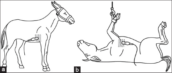
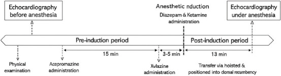
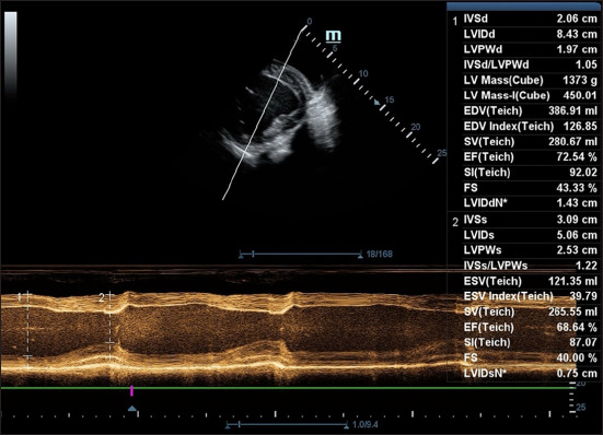
Materials and methods: Six healthy adult mules (18.83 ± 0.75 years; 263.83 ± 39.34 kg) were evaluated using standard two-dimensional and M-mode transthoracic echocardiography. Measurements were obtained before sedation (standing) and 13-min post-anesthetic induction (dorsal recumbency). Each mule received an intravenous injection of acepromazine (0.04 mg/kg), xylazine (1.6 mg/kg), diazepam (0.1 mg/kg), and ketamine (2.2 mg/kg). Key echocardiographic parameters included interventricular septum thickness (interventricular septum in diastole and interventricular septum in systole), left ventricular internal diameters (left ventricular internal diameter in diastole and left ventricular internal diameter in systole [LVIDs]), posterior wall thickness (left ventricular posterior wall in diastole and left ventricular posterior wall in systole), ejection fraction (EF), and fractional shortening (FS). Statistical comparisons were made using paired t-tests and Wilcoxon signed-rank tests (p < 0.05).
Results: Heart rate, EF, and FS significantly decreased post-anesthesia (p < 0.01), indicating reduced systolic function. Specifically, LVIDs increased from 4.60 ± 0.65 cm to 6.26 ± 0.48 cm (p < 0.01), while no significant changes were observed in diastolic parameters or respiratory rate. Anesthetic induction was smooth and graded as good to excellent in all cases.
Conclusion: High-dose xylazine significantly suppressed systolic cardiac function in anesthetized mules without causing arrhythmias or bradyarrhythmia. The combination protocol was effective and provided safe anesthesia induction, with echocardiography proving feasible under dorsal recumbency. These findings support the cautious use of upper-limit xylazine dosing in mules and suggest echocardiographic monitoring as a valuable tool during anesthesia.
期刊介绍:
Veterinary World publishes high quality papers focusing on Veterinary and Animal Science. The fields of study are bacteriology, parasitology, pathology, virology, immunology, mycology, public health, biotechnology, meat science, fish diseases, nutrition, gynecology, genetics, wildlife, laboratory animals, animal models of human infections, prion diseases and epidemiology. Studies on zoonotic and emerging infections are highly appreciated. Review articles are highly appreciated. All articles published by Veterinary World are made freely and permanently accessible online. All articles to Veterinary World are posted online immediately as they are ready for publication.

 求助内容:
求助内容: 应助结果提醒方式:


