{"title":"胰腺血管母细胞瘤合并希佩尔-林道病1例","authors":"Naoto Nakamura, Yosuke Kasai, Kazuyuki Nagai, Asahi Sato, Kentaro Kadono, Norimitsu Uza, Tsuyoshi Ohno, Sho Koyasu, Yuji Nakamoto, Noritaka Sano, Ayako Takahashi, Shinya Otsuki, Hiroaki Ito, Kei Yamane, Takayuki Anazawa, Satoshi Ogiso, Yoichiro Uchida, Takashi Ito, Takamichi Ishii, Etsuro Hatano","doi":"10.70352/scrj.cr.25-0247","DOIUrl":null,"url":null,"abstract":"<p><strong>Introduction: </strong>von Hippel-Lindau (VHL) disease is an autosomal dominant hereditary disorder characterized by the development of tumor-like lesions in multiple organs. While central nervous system hemangioblastomas, pancreatic neuroendocrine tumors, and pancreatic cysts are commonly associated with VHL disease, there have been few reported cases of pancreatic hemangioblastoma in patients with VHL disease.</p><p><strong>Case presentation: </strong>A male patient in his 30s had been diagnosed with VHL disease and had been followed for cerebellar and spinal hemangioblastomas, and renal cell carcinoma, for which he had undergone several tumor resections, radiation therapy, and a ventriculoperitoneal shunt. A pancreatic head tumor deemed to be a neuroendocrine tumor on imaging findings exhibited a gradual increase in size from 12 to 33 mm for the past 2 years, but it had been monitored due to his comorbidities and declining daily living activities. Severe anemia was detected during his regular outpatient visit, and an emergency esophagogastroduodenoscopy revealed a submucosal tumor near the duodenal papilla with ulceration and active bleeding, making endoscopic hemostasis challenging. Dynamic contrast-enhanced CT showed active bleeding from the pancreatic tumor. Subsequently, emergency angiography was performed via the superior mesenteric artery, successfully embolizing vessels supplied by the inferior pancreaticoduodenal artery to achieve hemostasis. Due to concerns about rebleeding, we performed pancreaticoduodenectomy 1 month after the emergency angiography, during which we awaited the improvement of the patient's overall condition. Microscopic findings of the tumor showed multinodular proliferation with hematoxylin-eosin staining, revealing cells with clear cytoplasm and abundant capillaries and dilated branching vessels within the nests. Immunohistochemical analysis demonstrated positivity for alpha-inhibin and S100, with partial positivity for carbonic anhydrase IX, leading to a diagnosis of pancreatic hemangioblastoma.</p><p><strong>Conclusions: </strong>This paper reports a rare case of pancreatic hemangioblastoma arising in a patient with VHL disease. It is crucial to consider the possibility of pancreatic hemangioblastoma when treating pancreatic tumors in VHL disease patients.</p>","PeriodicalId":22096,"journal":{"name":"Surgical Case Reports","volume":"11 1","pages":""},"PeriodicalIF":0.7000,"publicationDate":"2025-01-01","publicationTypes":"Journal Article","fieldsOfStudy":null,"isOpenAccess":false,"openAccessPdf":"https://www.ncbi.nlm.nih.gov/pmc/articles/PMC12415619/pdf/","citationCount":"0","resultStr":"{\"title\":\"Pancreatic Hemangioblastoma in a Patient with von Hippel-Lindau Disease: A Case Report.\",\"authors\":\"Naoto Nakamura, Yosuke Kasai, Kazuyuki Nagai, Asahi Sato, Kentaro Kadono, Norimitsu Uza, Tsuyoshi Ohno, Sho Koyasu, Yuji Nakamoto, Noritaka Sano, Ayako Takahashi, Shinya Otsuki, Hiroaki Ito, Kei Yamane, Takayuki Anazawa, Satoshi Ogiso, Yoichiro Uchida, Takashi Ito, Takamichi Ishii, Etsuro Hatano\",\"doi\":\"10.70352/scrj.cr.25-0247\",\"DOIUrl\":null,\"url\":null,\"abstract\":\"<p><strong>Introduction: </strong>von Hippel-Lindau (VHL) disease is an autosomal dominant hereditary disorder characterized by the development of tumor-like lesions in multiple organs. While central nervous system hemangioblastomas, pancreatic neuroendocrine tumors, and pancreatic cysts are commonly associated with VHL disease, there have been few reported cases of pancreatic hemangioblastoma in patients with VHL disease.</p><p><strong>Case presentation: </strong>A male patient in his 30s had been diagnosed with VHL disease and had been followed for cerebellar and spinal hemangioblastomas, and renal cell carcinoma, for which he had undergone several tumor resections, radiation therapy, and a ventriculoperitoneal shunt. A pancreatic head tumor deemed to be a neuroendocrine tumor on imaging findings exhibited a gradual increase in size from 12 to 33 mm for the past 2 years, but it had been monitored due to his comorbidities and declining daily living activities. Severe anemia was detected during his regular outpatient visit, and an emergency esophagogastroduodenoscopy revealed a submucosal tumor near the duodenal papilla with ulceration and active bleeding, making endoscopic hemostasis challenging. Dynamic contrast-enhanced CT showed active bleeding from the pancreatic tumor. Subsequently, emergency angiography was performed via the superior mesenteric artery, successfully embolizing vessels supplied by the inferior pancreaticoduodenal artery to achieve hemostasis. Due to concerns about rebleeding, we performed pancreaticoduodenectomy 1 month after the emergency angiography, during which we awaited the improvement of the patient's overall condition. Microscopic findings of the tumor showed multinodular proliferation with hematoxylin-eosin staining, revealing cells with clear cytoplasm and abundant capillaries and dilated branching vessels within the nests. Immunohistochemical analysis demonstrated positivity for alpha-inhibin and S100, with partial positivity for carbonic anhydrase IX, leading to a diagnosis of pancreatic hemangioblastoma.</p><p><strong>Conclusions: </strong>This paper reports a rare case of pancreatic hemangioblastoma arising in a patient with VHL disease. It is crucial to consider the possibility of pancreatic hemangioblastoma when treating pancreatic tumors in VHL disease patients.</p>\",\"PeriodicalId\":22096,\"journal\":{\"name\":\"Surgical Case Reports\",\"volume\":\"11 1\",\"pages\":\"\"},\"PeriodicalIF\":0.7000,\"publicationDate\":\"2025-01-01\",\"publicationTypes\":\"Journal Article\",\"fieldsOfStudy\":null,\"isOpenAccess\":false,\"openAccessPdf\":\"https://www.ncbi.nlm.nih.gov/pmc/articles/PMC12415619/pdf/\",\"citationCount\":\"0\",\"resultStr\":null,\"platform\":\"Semanticscholar\",\"paperid\":null,\"PeriodicalName\":\"Surgical Case Reports\",\"FirstCategoryId\":\"1085\",\"ListUrlMain\":\"https://doi.org/10.70352/scrj.cr.25-0247\",\"RegionNum\":0,\"RegionCategory\":null,\"ArticlePicture\":[],\"TitleCN\":null,\"AbstractTextCN\":null,\"PMCID\":null,\"EPubDate\":\"2025/9/5 0:00:00\",\"PubModel\":\"Epub\",\"JCR\":\"Q4\",\"JCRName\":\"SURGERY\",\"Score\":null,\"Total\":0}","platform":"Semanticscholar","paperid":null,"PeriodicalName":"Surgical Case Reports","FirstCategoryId":"1085","ListUrlMain":"https://doi.org/10.70352/scrj.cr.25-0247","RegionNum":0,"RegionCategory":null,"ArticlePicture":[],"TitleCN":null,"AbstractTextCN":null,"PMCID":null,"EPubDate":"2025/9/5 0:00:00","PubModel":"Epub","JCR":"Q4","JCRName":"SURGERY","Score":null,"Total":0}
Pancreatic Hemangioblastoma in a Patient with von Hippel-Lindau Disease: A Case Report.
Introduction: von Hippel-Lindau (VHL) disease is an autosomal dominant hereditary disorder characterized by the development of tumor-like lesions in multiple organs. While central nervous system hemangioblastomas, pancreatic neuroendocrine tumors, and pancreatic cysts are commonly associated with VHL disease, there have been few reported cases of pancreatic hemangioblastoma in patients with VHL disease.
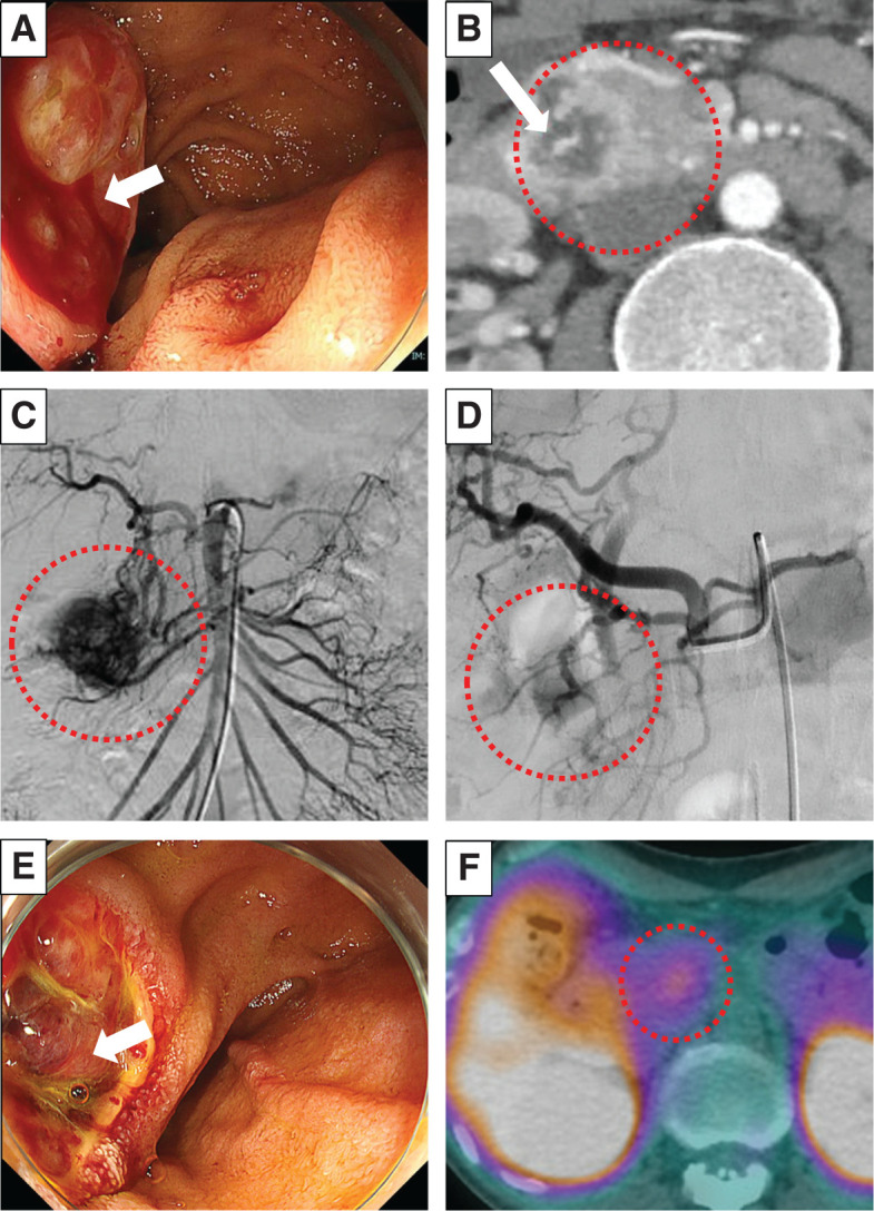
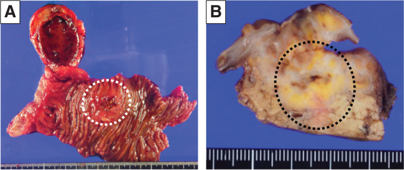
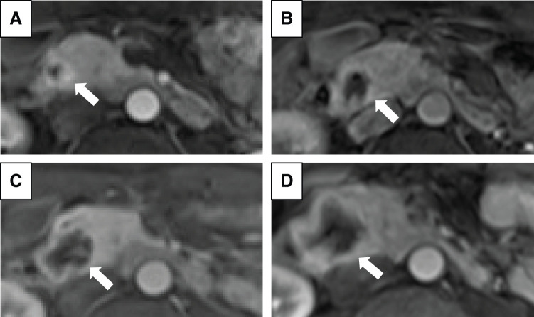
Case presentation: A male patient in his 30s had been diagnosed with VHL disease and had been followed for cerebellar and spinal hemangioblastomas, and renal cell carcinoma, for which he had undergone several tumor resections, radiation therapy, and a ventriculoperitoneal shunt. A pancreatic head tumor deemed to be a neuroendocrine tumor on imaging findings exhibited a gradual increase in size from 12 to 33 mm for the past 2 years, but it had been monitored due to his comorbidities and declining daily living activities. Severe anemia was detected during his regular outpatient visit, and an emergency esophagogastroduodenoscopy revealed a submucosal tumor near the duodenal papilla with ulceration and active bleeding, making endoscopic hemostasis challenging. Dynamic contrast-enhanced CT showed active bleeding from the pancreatic tumor. Subsequently, emergency angiography was performed via the superior mesenteric artery, successfully embolizing vessels supplied by the inferior pancreaticoduodenal artery to achieve hemostasis. Due to concerns about rebleeding, we performed pancreaticoduodenectomy 1 month after the emergency angiography, during which we awaited the improvement of the patient's overall condition. Microscopic findings of the tumor showed multinodular proliferation with hematoxylin-eosin staining, revealing cells with clear cytoplasm and abundant capillaries and dilated branching vessels within the nests. Immunohistochemical analysis demonstrated positivity for alpha-inhibin and S100, with partial positivity for carbonic anhydrase IX, leading to a diagnosis of pancreatic hemangioblastoma.
Conclusions: This paper reports a rare case of pancreatic hemangioblastoma arising in a patient with VHL disease. It is crucial to consider the possibility of pancreatic hemangioblastoma when treating pancreatic tumors in VHL disease patients.

 求助内容:
求助内容: 应助结果提醒方式:


