Marco Ninghetto, Georgios A Keliris, Kamil Szulborski, Tomasz Gałecki, Bartosz Kossowski, Daan Panneman, Frans P M Cremers, Monika Ołdak, Jacek P Szaflik, Kalina Burnat
{"title":"皮层对短暂和长期视野丧失的反应。","authors":"Marco Ninghetto, Georgios A Keliris, Kamil Szulborski, Tomasz Gałecki, Bartosz Kossowski, Daan Panneman, Frans P M Cremers, Monika Ołdak, Jacek P Szaflik, Kalina Burnat","doi":"10.1093/cercor/bhaf237","DOIUrl":null,"url":null,"abstract":"<p><p>In the visual cortices, receptive fields (RFs) are arranged in a gradient from small sizes in the center of the visual field to the largest sizes at the periphery. Using functional magnetic resonance imaging (fMRI) mapping of population RFs, we investigated RF adaptation in V1, V2, and V3 in patients after long-term photoreceptor degeneration affecting the central (Stargardt disease [STGD]) and peripheral (Retinitis Pigmentosa [RP]) regions of the retina. In controls, we temporarily limited the visual field to the central 10° to model peripheral loss. The central loss experienced by STGD patients led to an increase in RF size in the dorsal subdivisions of V1, V2, and V3. In contrast, peripheral loss in RP patients led to a bilateral increase in population RF sizes in V1 but a decrease in V2. Transient peripheral loss in controls led to an increase in RF size in V1 and a decrease in V2 and V3, regardless of the dorsal-ventral division of the cortical representation. Our findings suggest a dorsal-ventral difference in RF size in response to central visual field loss, likely reflecting the functional relevance of these divisions within the cortical representations of the visual field.</p>","PeriodicalId":9715,"journal":{"name":"Cerebral cortex","volume":"35 8","pages":""},"PeriodicalIF":2.9000,"publicationDate":"2025-08-01","publicationTypes":"Journal Article","fieldsOfStudy":null,"isOpenAccess":false,"openAccessPdf":"https://www.ncbi.nlm.nih.gov/pmc/articles/PMC12418949/pdf/","citationCount":"0","resultStr":"{\"title\":\"Cortical response to transient and long-term visual field loss.\",\"authors\":\"Marco Ninghetto, Georgios A Keliris, Kamil Szulborski, Tomasz Gałecki, Bartosz Kossowski, Daan Panneman, Frans P M Cremers, Monika Ołdak, Jacek P Szaflik, Kalina Burnat\",\"doi\":\"10.1093/cercor/bhaf237\",\"DOIUrl\":null,\"url\":null,\"abstract\":\"<p><p>In the visual cortices, receptive fields (RFs) are arranged in a gradient from small sizes in the center of the visual field to the largest sizes at the periphery. Using functional magnetic resonance imaging (fMRI) mapping of population RFs, we investigated RF adaptation in V1, V2, and V3 in patients after long-term photoreceptor degeneration affecting the central (Stargardt disease [STGD]) and peripheral (Retinitis Pigmentosa [RP]) regions of the retina. In controls, we temporarily limited the visual field to the central 10° to model peripheral loss. The central loss experienced by STGD patients led to an increase in RF size in the dorsal subdivisions of V1, V2, and V3. In contrast, peripheral loss in RP patients led to a bilateral increase in population RF sizes in V1 but a decrease in V2. Transient peripheral loss in controls led to an increase in RF size in V1 and a decrease in V2 and V3, regardless of the dorsal-ventral division of the cortical representation. Our findings suggest a dorsal-ventral difference in RF size in response to central visual field loss, likely reflecting the functional relevance of these divisions within the cortical representations of the visual field.</p>\",\"PeriodicalId\":9715,\"journal\":{\"name\":\"Cerebral cortex\",\"volume\":\"35 8\",\"pages\":\"\"},\"PeriodicalIF\":2.9000,\"publicationDate\":\"2025-08-01\",\"publicationTypes\":\"Journal Article\",\"fieldsOfStudy\":null,\"isOpenAccess\":false,\"openAccessPdf\":\"https://www.ncbi.nlm.nih.gov/pmc/articles/PMC12418949/pdf/\",\"citationCount\":\"0\",\"resultStr\":null,\"platform\":\"Semanticscholar\",\"paperid\":null,\"PeriodicalName\":\"Cerebral cortex\",\"FirstCategoryId\":\"3\",\"ListUrlMain\":\"https://doi.org/10.1093/cercor/bhaf237\",\"RegionNum\":2,\"RegionCategory\":\"医学\",\"ArticlePicture\":[],\"TitleCN\":null,\"AbstractTextCN\":null,\"PMCID\":null,\"EPubDate\":\"\",\"PubModel\":\"\",\"JCR\":\"Q2\",\"JCRName\":\"NEUROSCIENCES\",\"Score\":null,\"Total\":0}","platform":"Semanticscholar","paperid":null,"PeriodicalName":"Cerebral cortex","FirstCategoryId":"3","ListUrlMain":"https://doi.org/10.1093/cercor/bhaf237","RegionNum":2,"RegionCategory":"医学","ArticlePicture":[],"TitleCN":null,"AbstractTextCN":null,"PMCID":null,"EPubDate":"","PubModel":"","JCR":"Q2","JCRName":"NEUROSCIENCES","Score":null,"Total":0}
Cortical response to transient and long-term visual field loss.
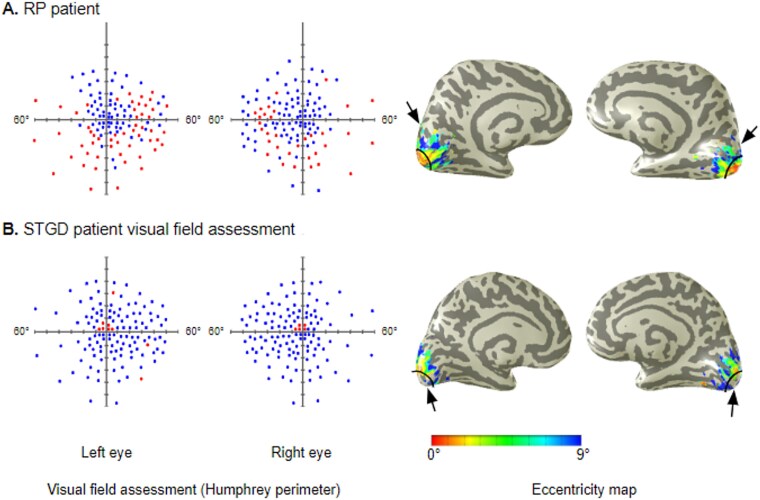
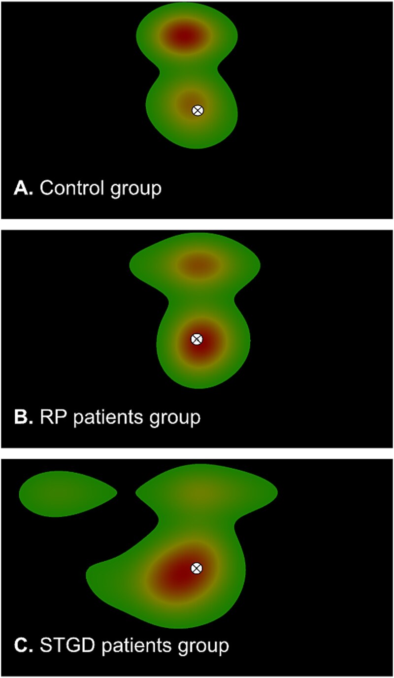
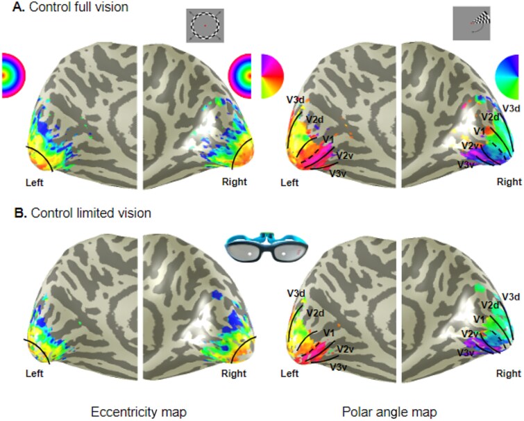
In the visual cortices, receptive fields (RFs) are arranged in a gradient from small sizes in the center of the visual field to the largest sizes at the periphery. Using functional magnetic resonance imaging (fMRI) mapping of population RFs, we investigated RF adaptation in V1, V2, and V3 in patients after long-term photoreceptor degeneration affecting the central (Stargardt disease [STGD]) and peripheral (Retinitis Pigmentosa [RP]) regions of the retina. In controls, we temporarily limited the visual field to the central 10° to model peripheral loss. The central loss experienced by STGD patients led to an increase in RF size in the dorsal subdivisions of V1, V2, and V3. In contrast, peripheral loss in RP patients led to a bilateral increase in population RF sizes in V1 but a decrease in V2. Transient peripheral loss in controls led to an increase in RF size in V1 and a decrease in V2 and V3, regardless of the dorsal-ventral division of the cortical representation. Our findings suggest a dorsal-ventral difference in RF size in response to central visual field loss, likely reflecting the functional relevance of these divisions within the cortical representations of the visual field.
期刊介绍:
Cerebral Cortex publishes papers on the development, organization, plasticity, and function of the cerebral cortex, including the hippocampus. Studies with clear relevance to the cerebral cortex, such as the thalamocortical relationship or cortico-subcortical interactions, are also included.
The journal is multidisciplinary and covers the large variety of modern neurobiological and neuropsychological techniques, including anatomy, biochemistry, molecular neurobiology, electrophysiology, behavior, artificial intelligence, and theoretical modeling. In addition to research articles, special features such as brief reviews, book reviews, and commentaries are included.
 求助内容:
求助内容: 应助结果提醒方式:


