{"title":"Mg-Ca-Si-Zr纳米颗粒在变革性骨科治疗中的界面响应","authors":"Priya Singh, Somesh Agrawal, Deepak Khare, Vinod Tiwari and Ashutosh Kumar Dubey","doi":"10.1039/D5LF00045A","DOIUrl":null,"url":null,"abstract":"<p >Debris particles, discharged due to degradation and wear, initiate an inflammatory response at the implantation site or lead to aseptic loosening of the prosthesis, ultimately resulting in implant failure over time. The toxicity concern becomes more severe with the release of nano-sized debris particles due to augmented interfacial interactions, even if the bulk counterpart is highly biocompatible. From this perspective, the present study aims to assess the <em>in vivo</em> toxicity, both local and systemic, of Mg<small><sub>1−<em>x</em></sub></small>Ca<small><sub><em>x</em></sub></small>Si<small><sub>1−<em>x</em></sub></small>Zr<small><sub><em>x</em></sub></small>O<small><sub>3</sub></small> (<em>x</em> = 0–0.4) [MCSZO-<em>X</em>, <em>X</em> = 0–4] nanoparticles using a rat model. Initially, the <em>in vitro</em> cytotoxicity of varying concentrations (0.25, 2.5, and 25 mg ml<small><sup>−1</sup></small>) of MCSZO-<em>X</em> nanoparticles was evaluated using MG-63 cells. Cell proliferation increases after the early interfacial interactions. Following this, 100 μl of MCSZO nanoparticles (25 mg ml<small><sup>−1</sup></small>) was administered through intra-articular injection into the knee joint of male Wistar rats. Biochemical analyses revealed no pathological changes in the liver and kidney of the injected group of rats. Additionally, the histopathological analyses demonstrated that there is no inflammation resulting from interfacial interactions with injected nanoparticles in various organs such as the liver, heart, kidney and knee. Overall, these findings pave the way for further advancement in bone repair and implant design.</p>","PeriodicalId":101138,"journal":{"name":"RSC Applied Interfaces","volume":" 5","pages":" 1331-1344"},"PeriodicalIF":0.0000,"publicationDate":"2025-05-29","publicationTypes":"Journal Article","fieldsOfStudy":null,"isOpenAccess":false,"openAccessPdf":"https://pubs.rsc.org/en/content/articlepdf/2025/lf/d5lf00045a?page=search","citationCount":"0","resultStr":"{\"title\":\"Interfacial response of Mg–Ca–Si–Zr nanoparticles for transformative orthopedic therapeutics\",\"authors\":\"Priya Singh, Somesh Agrawal, Deepak Khare, Vinod Tiwari and Ashutosh Kumar Dubey\",\"doi\":\"10.1039/D5LF00045A\",\"DOIUrl\":null,\"url\":null,\"abstract\":\"<p >Debris particles, discharged due to degradation and wear, initiate an inflammatory response at the implantation site or lead to aseptic loosening of the prosthesis, ultimately resulting in implant failure over time. The toxicity concern becomes more severe with the release of nano-sized debris particles due to augmented interfacial interactions, even if the bulk counterpart is highly biocompatible. From this perspective, the present study aims to assess the <em>in vivo</em> toxicity, both local and systemic, of Mg<small><sub>1−<em>x</em></sub></small>Ca<small><sub><em>x</em></sub></small>Si<small><sub>1−<em>x</em></sub></small>Zr<small><sub><em>x</em></sub></small>O<small><sub>3</sub></small> (<em>x</em> = 0–0.4) [MCSZO-<em>X</em>, <em>X</em> = 0–4] nanoparticles using a rat model. Initially, the <em>in vitro</em> cytotoxicity of varying concentrations (0.25, 2.5, and 25 mg ml<small><sup>−1</sup></small>) of MCSZO-<em>X</em> nanoparticles was evaluated using MG-63 cells. Cell proliferation increases after the early interfacial interactions. Following this, 100 μl of MCSZO nanoparticles (25 mg ml<small><sup>−1</sup></small>) was administered through intra-articular injection into the knee joint of male Wistar rats. Biochemical analyses revealed no pathological changes in the liver and kidney of the injected group of rats. Additionally, the histopathological analyses demonstrated that there is no inflammation resulting from interfacial interactions with injected nanoparticles in various organs such as the liver, heart, kidney and knee. Overall, these findings pave the way for further advancement in bone repair and implant design.</p>\",\"PeriodicalId\":101138,\"journal\":{\"name\":\"RSC Applied Interfaces\",\"volume\":\" 5\",\"pages\":\" 1331-1344\"},\"PeriodicalIF\":0.0000,\"publicationDate\":\"2025-05-29\",\"publicationTypes\":\"Journal Article\",\"fieldsOfStudy\":null,\"isOpenAccess\":false,\"openAccessPdf\":\"https://pubs.rsc.org/en/content/articlepdf/2025/lf/d5lf00045a?page=search\",\"citationCount\":\"0\",\"resultStr\":null,\"platform\":\"Semanticscholar\",\"paperid\":null,\"PeriodicalName\":\"RSC Applied Interfaces\",\"FirstCategoryId\":\"1085\",\"ListUrlMain\":\"https://pubs.rsc.org/en/content/articlelanding/2025/lf/d5lf00045a\",\"RegionNum\":0,\"RegionCategory\":null,\"ArticlePicture\":[],\"TitleCN\":null,\"AbstractTextCN\":null,\"PMCID\":null,\"EPubDate\":\"\",\"PubModel\":\"\",\"JCR\":\"\",\"JCRName\":\"\",\"Score\":null,\"Total\":0}","platform":"Semanticscholar","paperid":null,"PeriodicalName":"RSC Applied Interfaces","FirstCategoryId":"1085","ListUrlMain":"https://pubs.rsc.org/en/content/articlelanding/2025/lf/d5lf00045a","RegionNum":0,"RegionCategory":null,"ArticlePicture":[],"TitleCN":null,"AbstractTextCN":null,"PMCID":null,"EPubDate":"","PubModel":"","JCR":"","JCRName":"","Score":null,"Total":0}
引用次数: 0
摘要
由于降解和磨损而排出的碎片颗粒会在植入部位引发炎症反应或导致假体无菌性松动,最终导致植入物失效。由于界面相互作用的增强,纳米级碎片颗粒的释放会使毒性问题变得更加严重,即使大块的对应物具有高度的生物相容性。从这个角度来看,本研究旨在通过大鼠模型评估Mg1−xCaxSi1−xZrxO3 (x = 0-0.4) [MCSZO-X, x = 0-4]纳米颗粒的体内局部和全身毒性。最初,使用mg -63细胞评估不同浓度(0.25、2.5和25 mg ml - 1)的MCSZO-X纳米颗粒的体外细胞毒性。早期界面相互作用后细胞增殖增加。随后,将MCSZO纳米颗粒(25 mg ml−1)100 μl通过关节内注射至雄性Wistar大鼠膝关节。生化分析显示注射组大鼠肝脏和肾脏未见病理改变。此外,组织病理学分析表明,在肝脏、心脏、肾脏和膝盖等各种器官中,注射的纳米颗粒与界面相互作用没有引起炎症。总的来说,这些发现为骨修复和种植体设计的进一步发展铺平了道路。
Interfacial response of Mg–Ca–Si–Zr nanoparticles for transformative orthopedic therapeutics
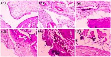
Debris particles, discharged due to degradation and wear, initiate an inflammatory response at the implantation site or lead to aseptic loosening of the prosthesis, ultimately resulting in implant failure over time. The toxicity concern becomes more severe with the release of nano-sized debris particles due to augmented interfacial interactions, even if the bulk counterpart is highly biocompatible. From this perspective, the present study aims to assess the in vivo toxicity, both local and systemic, of Mg1−xCaxSi1−xZrxO3 (x = 0–0.4) [MCSZO-X, X = 0–4] nanoparticles using a rat model. Initially, the in vitro cytotoxicity of varying concentrations (0.25, 2.5, and 25 mg ml−1) of MCSZO-X nanoparticles was evaluated using MG-63 cells. Cell proliferation increases after the early interfacial interactions. Following this, 100 μl of MCSZO nanoparticles (25 mg ml−1) was administered through intra-articular injection into the knee joint of male Wistar rats. Biochemical analyses revealed no pathological changes in the liver and kidney of the injected group of rats. Additionally, the histopathological analyses demonstrated that there is no inflammation resulting from interfacial interactions with injected nanoparticles in various organs such as the liver, heart, kidney and knee. Overall, these findings pave the way for further advancement in bone repair and implant design.

 求助内容:
求助内容: 应助结果提醒方式:


