{"title":"左心室整体纵向应变在采集和分析技术上的可靠性:一项前瞻性比较研究。","authors":"FeiFei Gong, Nausheen Akhter, Inga Vaitenas, Bernadette Wodzinski, Nicola Lancki, Leah J Welty, Kameswari Maganti","doi":"10.1093/ehjimp/qyaf101","DOIUrl":null,"url":null,"abstract":"<p><strong>Aims: </strong>Left ventricular (LV) global longitudinal strain (GLS) is a sensitive marker for detection of subclinical LV systolic dysfunction, but variability in acquisition and analysis may limit its clinical utility. We studied the accuracy, variability, and correlation of LV GLS across different 2D and 3D echocardiographic acquisition methods and post-processing platforms.</p><p><strong>Methods and results: </strong>In this prospective study, we analyzed 254 consecutive patients (mean age 55 ± 16 years, 60% female) undergoing clinically indicated echo. GLS was measured using multiple 2D acquisition methods (three beats and single beat) and 3D. Analyses were performed using both vendor-specific (EchoPac) and vendor-neutral (TomTec-Arena) software. Correlations and agreement between methods were assessed using Pearson correlation, intraclass correlation coefficients (ICCs), and Bland-Altman analyses.GLS values were highly consistent across the acquisition methods and between software platforms. Mean GLS values were -19.4 ± 3.3 (2D-A), -19.2 ± 3.3 (2D-B), -19.1 ± 3.5 (3P), and -14.8 ± 4.1 (3D). Intra- and interobserver variability for 2D GLS was low (ICC >0.9), indicating excellent reproducibility. However, 3D GLS values were significantly lower than 2D (mean difference -4.3%), with only moderate correlation (<i>r</i> = 0.66), suggesting that 2D and 3D GLS values are not interchangeable.</p><p><strong>Conclusion: </strong>The LV GLS is a reliable method for assessment of LV function with strong reproducibility across differing acquisition and analysis methods. However, 3D GLS is consistently lower and should not be used interchangeably with 2D measurements. These findings underscore the need for ongoing standardization and caution in comparing GLS values across 2D and 3D methods.</p>","PeriodicalId":94317,"journal":{"name":"European heart journal. Imaging methods and practice","volume":"3 2","pages":"qyaf101"},"PeriodicalIF":0.0000,"publicationDate":"2025-08-26","publicationTypes":"Journal Article","fieldsOfStudy":null,"isOpenAccess":false,"openAccessPdf":"https://www.ncbi.nlm.nih.gov/pmc/articles/PMC12405870/pdf/","citationCount":"0","resultStr":"{\"title\":\"Reliability of left ventricular global longitudinal strain across acquisition and analysis techniques: a prospective comparative study.\",\"authors\":\"FeiFei Gong, Nausheen Akhter, Inga Vaitenas, Bernadette Wodzinski, Nicola Lancki, Leah J Welty, Kameswari Maganti\",\"doi\":\"10.1093/ehjimp/qyaf101\",\"DOIUrl\":null,\"url\":null,\"abstract\":\"<p><strong>Aims: </strong>Left ventricular (LV) global longitudinal strain (GLS) is a sensitive marker for detection of subclinical LV systolic dysfunction, but variability in acquisition and analysis may limit its clinical utility. We studied the accuracy, variability, and correlation of LV GLS across different 2D and 3D echocardiographic acquisition methods and post-processing platforms.</p><p><strong>Methods and results: </strong>In this prospective study, we analyzed 254 consecutive patients (mean age 55 ± 16 years, 60% female) undergoing clinically indicated echo. GLS was measured using multiple 2D acquisition methods (three beats and single beat) and 3D. Analyses were performed using both vendor-specific (EchoPac) and vendor-neutral (TomTec-Arena) software. Correlations and agreement between methods were assessed using Pearson correlation, intraclass correlation coefficients (ICCs), and Bland-Altman analyses.GLS values were highly consistent across the acquisition methods and between software platforms. Mean GLS values were -19.4 ± 3.3 (2D-A), -19.2 ± 3.3 (2D-B), -19.1 ± 3.5 (3P), and -14.8 ± 4.1 (3D). Intra- and interobserver variability for 2D GLS was low (ICC >0.9), indicating excellent reproducibility. However, 3D GLS values were significantly lower than 2D (mean difference -4.3%), with only moderate correlation (<i>r</i> = 0.66), suggesting that 2D and 3D GLS values are not interchangeable.</p><p><strong>Conclusion: </strong>The LV GLS is a reliable method for assessment of LV function with strong reproducibility across differing acquisition and analysis methods. However, 3D GLS is consistently lower and should not be used interchangeably with 2D measurements. These findings underscore the need for ongoing standardization and caution in comparing GLS values across 2D and 3D methods.</p>\",\"PeriodicalId\":94317,\"journal\":{\"name\":\"European heart journal. Imaging methods and practice\",\"volume\":\"3 2\",\"pages\":\"qyaf101\"},\"PeriodicalIF\":0.0000,\"publicationDate\":\"2025-08-26\",\"publicationTypes\":\"Journal Article\",\"fieldsOfStudy\":null,\"isOpenAccess\":false,\"openAccessPdf\":\"https://www.ncbi.nlm.nih.gov/pmc/articles/PMC12405870/pdf/\",\"citationCount\":\"0\",\"resultStr\":null,\"platform\":\"Semanticscholar\",\"paperid\":null,\"PeriodicalName\":\"European heart journal. Imaging methods and practice\",\"FirstCategoryId\":\"1085\",\"ListUrlMain\":\"https://doi.org/10.1093/ehjimp/qyaf101\",\"RegionNum\":0,\"RegionCategory\":null,\"ArticlePicture\":[],\"TitleCN\":null,\"AbstractTextCN\":null,\"PMCID\":null,\"EPubDate\":\"2025/7/1 0:00:00\",\"PubModel\":\"eCollection\",\"JCR\":\"\",\"JCRName\":\"\",\"Score\":null,\"Total\":0}","platform":"Semanticscholar","paperid":null,"PeriodicalName":"European heart journal. Imaging methods and practice","FirstCategoryId":"1085","ListUrlMain":"https://doi.org/10.1093/ehjimp/qyaf101","RegionNum":0,"RegionCategory":null,"ArticlePicture":[],"TitleCN":null,"AbstractTextCN":null,"PMCID":null,"EPubDate":"2025/7/1 0:00:00","PubModel":"eCollection","JCR":"","JCRName":"","Score":null,"Total":0}
Reliability of left ventricular global longitudinal strain across acquisition and analysis techniques: a prospective comparative study.
Aims: Left ventricular (LV) global longitudinal strain (GLS) is a sensitive marker for detection of subclinical LV systolic dysfunction, but variability in acquisition and analysis may limit its clinical utility. We studied the accuracy, variability, and correlation of LV GLS across different 2D and 3D echocardiographic acquisition methods and post-processing platforms.
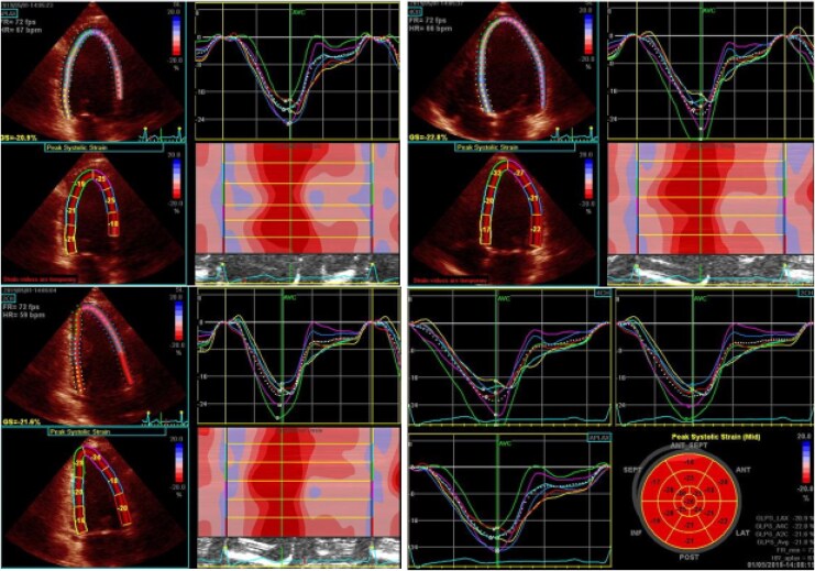
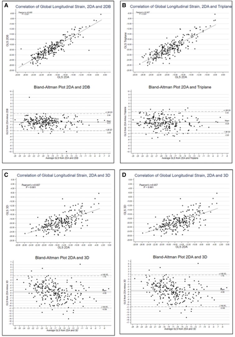
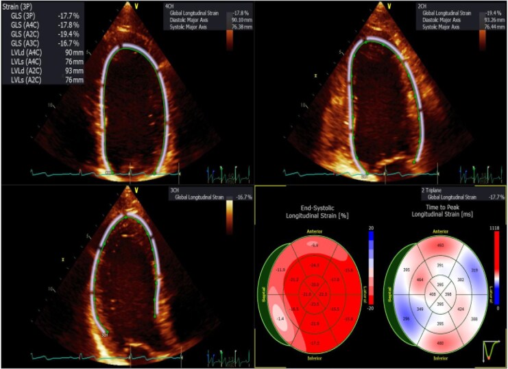
Methods and results: In this prospective study, we analyzed 254 consecutive patients (mean age 55 ± 16 years, 60% female) undergoing clinically indicated echo. GLS was measured using multiple 2D acquisition methods (three beats and single beat) and 3D. Analyses were performed using both vendor-specific (EchoPac) and vendor-neutral (TomTec-Arena) software. Correlations and agreement between methods were assessed using Pearson correlation, intraclass correlation coefficients (ICCs), and Bland-Altman analyses.GLS values were highly consistent across the acquisition methods and between software platforms. Mean GLS values were -19.4 ± 3.3 (2D-A), -19.2 ± 3.3 (2D-B), -19.1 ± 3.5 (3P), and -14.8 ± 4.1 (3D). Intra- and interobserver variability for 2D GLS was low (ICC >0.9), indicating excellent reproducibility. However, 3D GLS values were significantly lower than 2D (mean difference -4.3%), with only moderate correlation (r = 0.66), suggesting that 2D and 3D GLS values are not interchangeable.
Conclusion: The LV GLS is a reliable method for assessment of LV function with strong reproducibility across differing acquisition and analysis methods. However, 3D GLS is consistently lower and should not be used interchangeably with 2D measurements. These findings underscore the need for ongoing standardization and caution in comparing GLS values across 2D and 3D methods.

 求助内容:
求助内容: 应助结果提醒方式:


