{"title":"利用弥散张量成像和神经束造影对钩状束的放射学评价:技术考虑和临床意义的回顾。","authors":"Anna Stefańska, Sara Kierońska-Siwak","doi":"10.5114/pjr/206075","DOIUrl":null,"url":null,"abstract":"<p><p>Diffusion tensor imaging (DTI) and tractography are powerful non-invasive techniques for studying the human brain's white matter pathways. The uncinate fasciculus (UF) is a key frontotemporal tract involved in emotion regulation, memory, and language. Despite advancements, challenges persist in accurately mapping its structure and function due to methodological limitations in data acquisition and analysis. This review aims to provide a comprehensive overview of the strengths and limitations of DTI and tractography in studying the UF, focusing on its anatomy, data acquisition techniques, and associated neurological and psychiatric disorders. A systematic review of over 30 years of literature on UF was conducted, encompassing anatomical studies, DTI methodologies, and clinical applications. Studies involving both postmortem dissections and <i>in vivo</i> imaging were analysed, with particular attention to different DTI acquisition parameters, fibre tracking algorithms, and their impact on imaging accuracy. DTI has significantly improved our understanding of UF anatomy and its role in neurocognitive functions. However, methodological constraints such as low spatial resolution, crossing fibres, and inter-subject variability limit its precision. Advances in higher-field magnetic resonance imaging, improved diffusion models, and artificial intelligence-enhanced tractography offer promising solutions. UF abnormalities have been linked to various disorders, including schizophrenia, depression, autism spectrum disorders, and neurodegenerative diseases. While DTI and tractography are invaluable tools for studying the UF, their limitations necessitate cautious interpretation of results. Future research should focus on refining imaging techniques to enhance accuracy and clinical applicability, paving the way for better diagnostic and therapeutic strategies.</p>","PeriodicalId":94174,"journal":{"name":"Polish journal of radiology","volume":"90 ","pages":"e324-e344"},"PeriodicalIF":0.0000,"publicationDate":"2025-07-07","publicationTypes":"Journal Article","fieldsOfStudy":null,"isOpenAccess":false,"openAccessPdf":"https://www.ncbi.nlm.nih.gov/pmc/articles/PMC12403653/pdf/","citationCount":"0","resultStr":"{\"title\":\"Radiologic evaluation of the uncinate fasciculus using diffusion tensor imaging and tractography: review of technical considerations and clinical implications.\",\"authors\":\"Anna Stefańska, Sara Kierońska-Siwak\",\"doi\":\"10.5114/pjr/206075\",\"DOIUrl\":null,\"url\":null,\"abstract\":\"<p><p>Diffusion tensor imaging (DTI) and tractography are powerful non-invasive techniques for studying the human brain's white matter pathways. The uncinate fasciculus (UF) is a key frontotemporal tract involved in emotion regulation, memory, and language. Despite advancements, challenges persist in accurately mapping its structure and function due to methodological limitations in data acquisition and analysis. This review aims to provide a comprehensive overview of the strengths and limitations of DTI and tractography in studying the UF, focusing on its anatomy, data acquisition techniques, and associated neurological and psychiatric disorders. A systematic review of over 30 years of literature on UF was conducted, encompassing anatomical studies, DTI methodologies, and clinical applications. Studies involving both postmortem dissections and <i>in vivo</i> imaging were analysed, with particular attention to different DTI acquisition parameters, fibre tracking algorithms, and their impact on imaging accuracy. DTI has significantly improved our understanding of UF anatomy and its role in neurocognitive functions. However, methodological constraints such as low spatial resolution, crossing fibres, and inter-subject variability limit its precision. Advances in higher-field magnetic resonance imaging, improved diffusion models, and artificial intelligence-enhanced tractography offer promising solutions. UF abnormalities have been linked to various disorders, including schizophrenia, depression, autism spectrum disorders, and neurodegenerative diseases. While DTI and tractography are invaluable tools for studying the UF, their limitations necessitate cautious interpretation of results. Future research should focus on refining imaging techniques to enhance accuracy and clinical applicability, paving the way for better diagnostic and therapeutic strategies.</p>\",\"PeriodicalId\":94174,\"journal\":{\"name\":\"Polish journal of radiology\",\"volume\":\"90 \",\"pages\":\"e324-e344\"},\"PeriodicalIF\":0.0000,\"publicationDate\":\"2025-07-07\",\"publicationTypes\":\"Journal Article\",\"fieldsOfStudy\":null,\"isOpenAccess\":false,\"openAccessPdf\":\"https://www.ncbi.nlm.nih.gov/pmc/articles/PMC12403653/pdf/\",\"citationCount\":\"0\",\"resultStr\":null,\"platform\":\"Semanticscholar\",\"paperid\":null,\"PeriodicalName\":\"Polish journal of radiology\",\"FirstCategoryId\":\"1085\",\"ListUrlMain\":\"https://doi.org/10.5114/pjr/206075\",\"RegionNum\":0,\"RegionCategory\":null,\"ArticlePicture\":[],\"TitleCN\":null,\"AbstractTextCN\":null,\"PMCID\":null,\"EPubDate\":\"2025/1/1 0:00:00\",\"PubModel\":\"eCollection\",\"JCR\":\"\",\"JCRName\":\"\",\"Score\":null,\"Total\":0}","platform":"Semanticscholar","paperid":null,"PeriodicalName":"Polish journal of radiology","FirstCategoryId":"1085","ListUrlMain":"https://doi.org/10.5114/pjr/206075","RegionNum":0,"RegionCategory":null,"ArticlePicture":[],"TitleCN":null,"AbstractTextCN":null,"PMCID":null,"EPubDate":"2025/1/1 0:00:00","PubModel":"eCollection","JCR":"","JCRName":"","Score":null,"Total":0}
Radiologic evaluation of the uncinate fasciculus using diffusion tensor imaging and tractography: review of technical considerations and clinical implications.
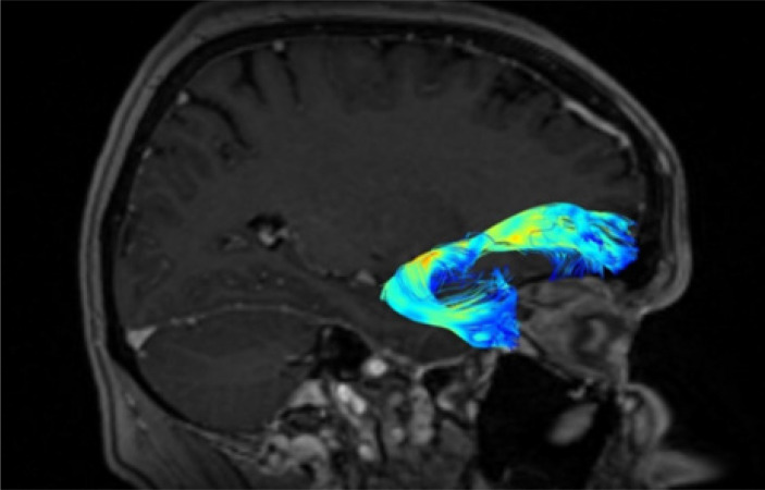
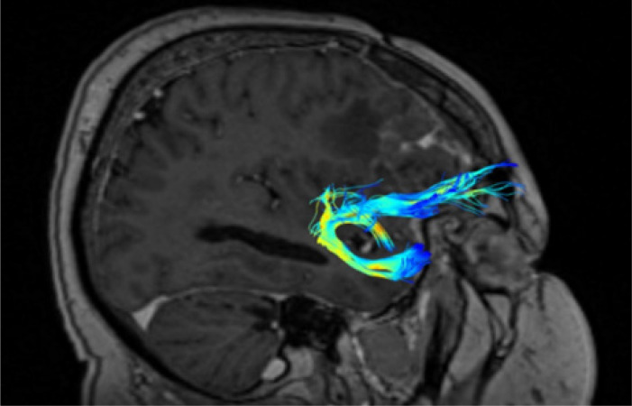
Diffusion tensor imaging (DTI) and tractography are powerful non-invasive techniques for studying the human brain's white matter pathways. The uncinate fasciculus (UF) is a key frontotemporal tract involved in emotion regulation, memory, and language. Despite advancements, challenges persist in accurately mapping its structure and function due to methodological limitations in data acquisition and analysis. This review aims to provide a comprehensive overview of the strengths and limitations of DTI and tractography in studying the UF, focusing on its anatomy, data acquisition techniques, and associated neurological and psychiatric disorders. A systematic review of over 30 years of literature on UF was conducted, encompassing anatomical studies, DTI methodologies, and clinical applications. Studies involving both postmortem dissections and in vivo imaging were analysed, with particular attention to different DTI acquisition parameters, fibre tracking algorithms, and their impact on imaging accuracy. DTI has significantly improved our understanding of UF anatomy and its role in neurocognitive functions. However, methodological constraints such as low spatial resolution, crossing fibres, and inter-subject variability limit its precision. Advances in higher-field magnetic resonance imaging, improved diffusion models, and artificial intelligence-enhanced tractography offer promising solutions. UF abnormalities have been linked to various disorders, including schizophrenia, depression, autism spectrum disorders, and neurodegenerative diseases. While DTI and tractography are invaluable tools for studying the UF, their limitations necessitate cautious interpretation of results. Future research should focus on refining imaging techniques to enhance accuracy and clinical applicability, paving the way for better diagnostic and therapeutic strategies.

 求助内容:
求助内容: 应助结果提醒方式:


