{"title":"角膜内皮失代偿和Descemet膜内皮角膜移植术的眼表改变。","authors":"Minglu Ma, Xiaojuan Dong, Jing Wu, Jiayin Peng, Ruilin Guo, Yi Yu, Chenjia Xu, Chen Ouyang, Ting Huang","doi":"10.2147/OPTH.S508390","DOIUrl":null,"url":null,"abstract":"<p><strong>Objective: </strong>To explore alterations in tears and ocular surface throughout Descemet's Membrane Endothelial Keratoplasty (DMEK), thereby offering valuable insights for clinical intervention.</p><p><strong>Methods: </strong>This study was conducted on 84 patients (84 eyes) with CED and received DMEK who visited the Zhongshan Ophthalmic Center of Sun Yat-sen University from April 2022 to January 2024. All patients were evaluated preoperatively and 1, 3, 6, and 12 months postoperatively, with 31 patients (31 eyes) post-24 months above. This cohort was then compared against a normal control of 21 individuals (21 eyes). Main clinical outcomes included lipid layer thickness (LLT), blink counts and partial blink rate (PBR) measured by Lipiview interferometer, in vivo confocal microscopy (IVCM) was used to detect the density of epithelial wing cells, leukocytes, subbasal dendritic cells (DCs) and endothelial cells.</p><p><strong>Results: </strong>Compared with normal control, LLT was decreased at 6 months (P = 0.053); total blink counts were significantly higher at 36 months (P < 0.05) after DMEK, while LLT was significantly lower (P < 0.05). Utilizing IVCM, an increased leukocytes density was identified in CED patients, which correlated positively with Log MAR visual acuity (r = 0.261, P = 0.04) and inversely with epithelial wing cells density (r = -0.319, P = 0.009). Leukocytes density decreased to comparable level at postoperative 3 months (P = 0.11). Subbasal DCs density was still higher at 6 months postoperatively (P < 0.05), while there was no difference at 12 months after DMEK (P > 0.05).</p><p><strong>Conclusion: </strong>Abnormalities of LLT can occur at different stages after DMEK. Besides, the immune reaction on cornea is still active within 6 months after DMEK.</p>","PeriodicalId":93945,"journal":{"name":"Clinical ophthalmology (Auckland, N.Z.)","volume":"19 ","pages":"2977-2984"},"PeriodicalIF":0.0000,"publicationDate":"2025-08-26","publicationTypes":"Journal Article","fieldsOfStudy":null,"isOpenAccess":false,"openAccessPdf":"https://www.ncbi.nlm.nih.gov/pmc/articles/PMC12399790/pdf/","citationCount":"0","resultStr":"{\"title\":\"Ocular Surface Alteration on Corneal Endothelial Decompensation and Descemet's Membrane Endothelial Keratoplasty.\",\"authors\":\"Minglu Ma, Xiaojuan Dong, Jing Wu, Jiayin Peng, Ruilin Guo, Yi Yu, Chenjia Xu, Chen Ouyang, Ting Huang\",\"doi\":\"10.2147/OPTH.S508390\",\"DOIUrl\":null,\"url\":null,\"abstract\":\"<p><strong>Objective: </strong>To explore alterations in tears and ocular surface throughout Descemet's Membrane Endothelial Keratoplasty (DMEK), thereby offering valuable insights for clinical intervention.</p><p><strong>Methods: </strong>This study was conducted on 84 patients (84 eyes) with CED and received DMEK who visited the Zhongshan Ophthalmic Center of Sun Yat-sen University from April 2022 to January 2024. All patients were evaluated preoperatively and 1, 3, 6, and 12 months postoperatively, with 31 patients (31 eyes) post-24 months above. This cohort was then compared against a normal control of 21 individuals (21 eyes). Main clinical outcomes included lipid layer thickness (LLT), blink counts and partial blink rate (PBR) measured by Lipiview interferometer, in vivo confocal microscopy (IVCM) was used to detect the density of epithelial wing cells, leukocytes, subbasal dendritic cells (DCs) and endothelial cells.</p><p><strong>Results: </strong>Compared with normal control, LLT was decreased at 6 months (P = 0.053); total blink counts were significantly higher at 36 months (P < 0.05) after DMEK, while LLT was significantly lower (P < 0.05). Utilizing IVCM, an increased leukocytes density was identified in CED patients, which correlated positively with Log MAR visual acuity (r = 0.261, P = 0.04) and inversely with epithelial wing cells density (r = -0.319, P = 0.009). Leukocytes density decreased to comparable level at postoperative 3 months (P = 0.11). Subbasal DCs density was still higher at 6 months postoperatively (P < 0.05), while there was no difference at 12 months after DMEK (P > 0.05).</p><p><strong>Conclusion: </strong>Abnormalities of LLT can occur at different stages after DMEK. Besides, the immune reaction on cornea is still active within 6 months after DMEK.</p>\",\"PeriodicalId\":93945,\"journal\":{\"name\":\"Clinical ophthalmology (Auckland, N.Z.)\",\"volume\":\"19 \",\"pages\":\"2977-2984\"},\"PeriodicalIF\":0.0000,\"publicationDate\":\"2025-08-26\",\"publicationTypes\":\"Journal Article\",\"fieldsOfStudy\":null,\"isOpenAccess\":false,\"openAccessPdf\":\"https://www.ncbi.nlm.nih.gov/pmc/articles/PMC12399790/pdf/\",\"citationCount\":\"0\",\"resultStr\":null,\"platform\":\"Semanticscholar\",\"paperid\":null,\"PeriodicalName\":\"Clinical ophthalmology (Auckland, N.Z.)\",\"FirstCategoryId\":\"1085\",\"ListUrlMain\":\"https://doi.org/10.2147/OPTH.S508390\",\"RegionNum\":0,\"RegionCategory\":null,\"ArticlePicture\":[],\"TitleCN\":null,\"AbstractTextCN\":null,\"PMCID\":null,\"EPubDate\":\"2025/1/1 0:00:00\",\"PubModel\":\"eCollection\",\"JCR\":\"\",\"JCRName\":\"\",\"Score\":null,\"Total\":0}","platform":"Semanticscholar","paperid":null,"PeriodicalName":"Clinical ophthalmology (Auckland, N.Z.)","FirstCategoryId":"1085","ListUrlMain":"https://doi.org/10.2147/OPTH.S508390","RegionNum":0,"RegionCategory":null,"ArticlePicture":[],"TitleCN":null,"AbstractTextCN":null,"PMCID":null,"EPubDate":"2025/1/1 0:00:00","PubModel":"eCollection","JCR":"","JCRName":"","Score":null,"Total":0}
Ocular Surface Alteration on Corneal Endothelial Decompensation and Descemet's Membrane Endothelial Keratoplasty.
Objective: To explore alterations in tears and ocular surface throughout Descemet's Membrane Endothelial Keratoplasty (DMEK), thereby offering valuable insights for clinical intervention.
Methods: This study was conducted on 84 patients (84 eyes) with CED and received DMEK who visited the Zhongshan Ophthalmic Center of Sun Yat-sen University from April 2022 to January 2024. All patients were evaluated preoperatively and 1, 3, 6, and 12 months postoperatively, with 31 patients (31 eyes) post-24 months above. This cohort was then compared against a normal control of 21 individuals (21 eyes). Main clinical outcomes included lipid layer thickness (LLT), blink counts and partial blink rate (PBR) measured by Lipiview interferometer, in vivo confocal microscopy (IVCM) was used to detect the density of epithelial wing cells, leukocytes, subbasal dendritic cells (DCs) and endothelial cells.
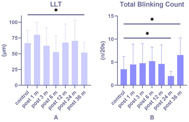
Results: Compared with normal control, LLT was decreased at 6 months (P = 0.053); total blink counts were significantly higher at 36 months (P < 0.05) after DMEK, while LLT was significantly lower (P < 0.05). Utilizing IVCM, an increased leukocytes density was identified in CED patients, which correlated positively with Log MAR visual acuity (r = 0.261, P = 0.04) and inversely with epithelial wing cells density (r = -0.319, P = 0.009). Leukocytes density decreased to comparable level at postoperative 3 months (P = 0.11). Subbasal DCs density was still higher at 6 months postoperatively (P < 0.05), while there was no difference at 12 months after DMEK (P > 0.05).
Conclusion: Abnormalities of LLT can occur at different stages after DMEK. Besides, the immune reaction on cornea is still active within 6 months after DMEK.


 求助内容:
求助内容: 应助结果提醒方式:


