{"title":"基于卷积神经网络和CBCT的牙龈厚度三维可视化与定量分析。","authors":"Lan Yang, ZiCheng Zhu, Yongshan Li, Jieying Huang, Xiaoli Wang, Haoran Zheng, Jiang Chen","doi":"10.3389/fdmed.2025.1635155","DOIUrl":null,"url":null,"abstract":"<p><strong>Objective: </strong>Traditional gingival thickness (GT) assessment methods provide only point measurements or simple classifications, lacking spatial distribution information. This study aimed to develop a CBCT-based 3D visualization system for gingival thickness using deep learning, providing a novel spatial assessment tool for implant surgery planning.</p><p><strong>Methods: </strong>CBCT and intraoral scanning (IOS) data from 50 patients with tooth loss were collected to establish a standardized dataset. DeepLabV3+ architecture was employed for semantic segmentation of gingival and bone tissues. A 3D visualization algorithm incorporating vertical scanning strategy, triangular mesh construction, and gradient color mapping was innovatively developed to transform 2D slices into continuous 3D surfaces.</p><p><strong>Results: </strong>The semantic segmentation model achieved a mIoU of 85.92 ± 0.43%. The 3D visualization system successfully constructed a comprehensive spatial distribution model of gingival thickness, clearly demonstrating GT variations from alveolar ridge to labial aspect through gradient coloration. The 3D model enabled millimeter-precision quantification, supporting multi-angle and multi-level GT assessment that overcame the limitations of traditional 2D measurements.</p><p><strong>Conclusion: </strong>This system represents a methodological advancement from qualitative to spatial quantitative GT assessment. The intuitive 3D visualization serves as an innovative preoperative tool that identifies high-risk areas and guides personalized surgical planning, enhancing predictability for aesthetic and complex implant cases.</p>","PeriodicalId":73077,"journal":{"name":"Frontiers in dental medicine","volume":"6 ","pages":"1635155"},"PeriodicalIF":1.8000,"publicationDate":"2025-08-18","publicationTypes":"Journal Article","fieldsOfStudy":null,"isOpenAccess":false,"openAccessPdf":"https://www.ncbi.nlm.nih.gov/pmc/articles/PMC12399636/pdf/","citationCount":"0","resultStr":"{\"title\":\"Clinical-oriented 3D visualization and quantitative analysis of gingival thickness using convolutional neural networks and CBCT.\",\"authors\":\"Lan Yang, ZiCheng Zhu, Yongshan Li, Jieying Huang, Xiaoli Wang, Haoran Zheng, Jiang Chen\",\"doi\":\"10.3389/fdmed.2025.1635155\",\"DOIUrl\":null,\"url\":null,\"abstract\":\"<p><strong>Objective: </strong>Traditional gingival thickness (GT) assessment methods provide only point measurements or simple classifications, lacking spatial distribution information. This study aimed to develop a CBCT-based 3D visualization system for gingival thickness using deep learning, providing a novel spatial assessment tool for implant surgery planning.</p><p><strong>Methods: </strong>CBCT and intraoral scanning (IOS) data from 50 patients with tooth loss were collected to establish a standardized dataset. DeepLabV3+ architecture was employed for semantic segmentation of gingival and bone tissues. A 3D visualization algorithm incorporating vertical scanning strategy, triangular mesh construction, and gradient color mapping was innovatively developed to transform 2D slices into continuous 3D surfaces.</p><p><strong>Results: </strong>The semantic segmentation model achieved a mIoU of 85.92 ± 0.43%. The 3D visualization system successfully constructed a comprehensive spatial distribution model of gingival thickness, clearly demonstrating GT variations from alveolar ridge to labial aspect through gradient coloration. The 3D model enabled millimeter-precision quantification, supporting multi-angle and multi-level GT assessment that overcame the limitations of traditional 2D measurements.</p><p><strong>Conclusion: </strong>This system represents a methodological advancement from qualitative to spatial quantitative GT assessment. The intuitive 3D visualization serves as an innovative preoperative tool that identifies high-risk areas and guides personalized surgical planning, enhancing predictability for aesthetic and complex implant cases.</p>\",\"PeriodicalId\":73077,\"journal\":{\"name\":\"Frontiers in dental medicine\",\"volume\":\"6 \",\"pages\":\"1635155\"},\"PeriodicalIF\":1.8000,\"publicationDate\":\"2025-08-18\",\"publicationTypes\":\"Journal Article\",\"fieldsOfStudy\":null,\"isOpenAccess\":false,\"openAccessPdf\":\"https://www.ncbi.nlm.nih.gov/pmc/articles/PMC12399636/pdf/\",\"citationCount\":\"0\",\"resultStr\":null,\"platform\":\"Semanticscholar\",\"paperid\":null,\"PeriodicalName\":\"Frontiers in dental medicine\",\"FirstCategoryId\":\"1085\",\"ListUrlMain\":\"https://doi.org/10.3389/fdmed.2025.1635155\",\"RegionNum\":0,\"RegionCategory\":null,\"ArticlePicture\":[],\"TitleCN\":null,\"AbstractTextCN\":null,\"PMCID\":null,\"EPubDate\":\"2025/1/1 0:00:00\",\"PubModel\":\"eCollection\",\"JCR\":\"Q3\",\"JCRName\":\"DENTISTRY, ORAL SURGERY & MEDICINE\",\"Score\":null,\"Total\":0}","platform":"Semanticscholar","paperid":null,"PeriodicalName":"Frontiers in dental medicine","FirstCategoryId":"1085","ListUrlMain":"https://doi.org/10.3389/fdmed.2025.1635155","RegionNum":0,"RegionCategory":null,"ArticlePicture":[],"TitleCN":null,"AbstractTextCN":null,"PMCID":null,"EPubDate":"2025/1/1 0:00:00","PubModel":"eCollection","JCR":"Q3","JCRName":"DENTISTRY, ORAL SURGERY & MEDICINE","Score":null,"Total":0}
Clinical-oriented 3D visualization and quantitative analysis of gingival thickness using convolutional neural networks and CBCT.
Objective: Traditional gingival thickness (GT) assessment methods provide only point measurements or simple classifications, lacking spatial distribution information. This study aimed to develop a CBCT-based 3D visualization system for gingival thickness using deep learning, providing a novel spatial assessment tool for implant surgery planning.
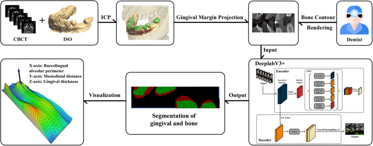
Methods: CBCT and intraoral scanning (IOS) data from 50 patients with tooth loss were collected to establish a standardized dataset. DeepLabV3+ architecture was employed for semantic segmentation of gingival and bone tissues. A 3D visualization algorithm incorporating vertical scanning strategy, triangular mesh construction, and gradient color mapping was innovatively developed to transform 2D slices into continuous 3D surfaces.
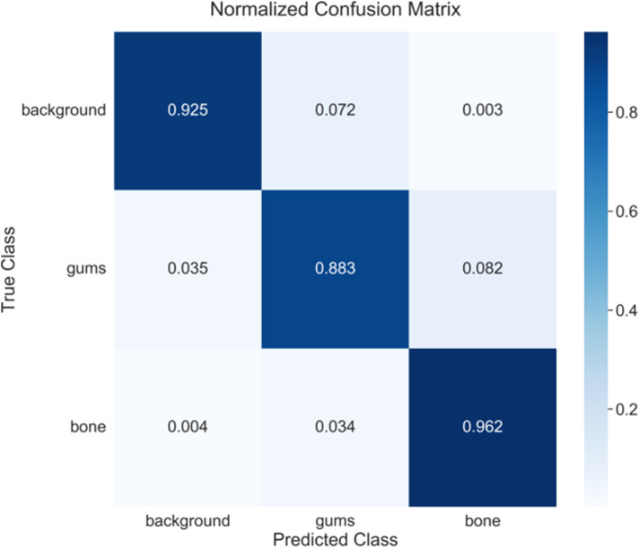
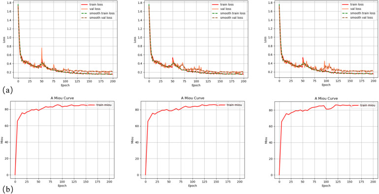
Results: The semantic segmentation model achieved a mIoU of 85.92 ± 0.43%. The 3D visualization system successfully constructed a comprehensive spatial distribution model of gingival thickness, clearly demonstrating GT variations from alveolar ridge to labial aspect through gradient coloration. The 3D model enabled millimeter-precision quantification, supporting multi-angle and multi-level GT assessment that overcame the limitations of traditional 2D measurements.
Conclusion: This system represents a methodological advancement from qualitative to spatial quantitative GT assessment. The intuitive 3D visualization serves as an innovative preoperative tool that identifies high-risk areas and guides personalized surgical planning, enhancing predictability for aesthetic and complex implant cases.

 求助内容:
求助内容: 应助结果提醒方式:


