Katelyn Cahill, Catriona Hargrave, Patrick O'Connor, Mark Denham, Nathan Hearn, Dinesh Vignarajah, Zack Y Shan, Myo Min
{"title":"利用表观扩散系数制定保甲状腺放射治疗计划的生物危险体积。","authors":"Katelyn Cahill, Catriona Hargrave, Patrick O'Connor, Mark Denham, Nathan Hearn, Dinesh Vignarajah, Zack Y Shan, Myo Min","doi":"10.1093/bjro/tzaf020","DOIUrl":null,"url":null,"abstract":"<p><strong>Objectives: </strong>Xerostomia toxicity continues to contribute towards a decrease in quality of life in head and neck cancer patients. Diffusion weighted MRI and the associated apparent diffusion coefficient (ADC) may identify the radiosensitive region within the parotid gland (PG). This study retrospectively assesses the feasibility of using percentile threshold values from the ADC map to generate a biological at-risk volume (BRV). The location and distribution of BRV are evaluated across the PG.</p><p><strong>Methods: </strong>Image registration between the planning CT and MRI-simulation images was performed and reviewed to ensure accurate translation of ADC data when contouring the PG. Histogram analysis was undertaken using the 20th, 30th, and 50th percentile ADC values of each contoured PG to form the BRV. The whole PG was split into 8 anatomical sectors at a common intersection point to evaluate the distribution of BRV throughout.</p><p><strong>Results: </strong>The BRV distribution for each percentile was mapped across the whole contoured PG and each anatomical sector contour. The largest distribution was predominantly found in the superolateral sectors.</p><p><strong>Conclusions: </strong>The 20th and 30th percentile ADC values can be used to form a BRV of the PG. The location of the BRV distribution indicates a potential relationship between ADC thresholds and the functional region of the PG.</p><p><strong>Advances in knowledge: </strong>The BRV is located in a favourable position within the PG and could be used to further spare this salivary gland during dose optimization. The feasibility of this approach will be explored in a future retrospective dosimetry study.</p>","PeriodicalId":72419,"journal":{"name":"BJR open","volume":"7 1","pages":"tzaf020"},"PeriodicalIF":2.1000,"publicationDate":"2025-08-13","publicationTypes":"Journal Article","fieldsOfStudy":null,"isOpenAccess":false,"openAccessPdf":"https://www.ncbi.nlm.nih.gov/pmc/articles/PMC12401576/pdf/","citationCount":"0","resultStr":"{\"title\":\"Development of a biological at-risk volume using apparent diffusion coefficient for parotid-sparing radiation therapy planning.\",\"authors\":\"Katelyn Cahill, Catriona Hargrave, Patrick O'Connor, Mark Denham, Nathan Hearn, Dinesh Vignarajah, Zack Y Shan, Myo Min\",\"doi\":\"10.1093/bjro/tzaf020\",\"DOIUrl\":null,\"url\":null,\"abstract\":\"<p><strong>Objectives: </strong>Xerostomia toxicity continues to contribute towards a decrease in quality of life in head and neck cancer patients. Diffusion weighted MRI and the associated apparent diffusion coefficient (ADC) may identify the radiosensitive region within the parotid gland (PG). This study retrospectively assesses the feasibility of using percentile threshold values from the ADC map to generate a biological at-risk volume (BRV). The location and distribution of BRV are evaluated across the PG.</p><p><strong>Methods: </strong>Image registration between the planning CT and MRI-simulation images was performed and reviewed to ensure accurate translation of ADC data when contouring the PG. Histogram analysis was undertaken using the 20th, 30th, and 50th percentile ADC values of each contoured PG to form the BRV. The whole PG was split into 8 anatomical sectors at a common intersection point to evaluate the distribution of BRV throughout.</p><p><strong>Results: </strong>The BRV distribution for each percentile was mapped across the whole contoured PG and each anatomical sector contour. The largest distribution was predominantly found in the superolateral sectors.</p><p><strong>Conclusions: </strong>The 20th and 30th percentile ADC values can be used to form a BRV of the PG. The location of the BRV distribution indicates a potential relationship between ADC thresholds and the functional region of the PG.</p><p><strong>Advances in knowledge: </strong>The BRV is located in a favourable position within the PG and could be used to further spare this salivary gland during dose optimization. The feasibility of this approach will be explored in a future retrospective dosimetry study.</p>\",\"PeriodicalId\":72419,\"journal\":{\"name\":\"BJR open\",\"volume\":\"7 1\",\"pages\":\"tzaf020\"},\"PeriodicalIF\":2.1000,\"publicationDate\":\"2025-08-13\",\"publicationTypes\":\"Journal Article\",\"fieldsOfStudy\":null,\"isOpenAccess\":false,\"openAccessPdf\":\"https://www.ncbi.nlm.nih.gov/pmc/articles/PMC12401576/pdf/\",\"citationCount\":\"0\",\"resultStr\":null,\"platform\":\"Semanticscholar\",\"paperid\":null,\"PeriodicalName\":\"BJR open\",\"FirstCategoryId\":\"1085\",\"ListUrlMain\":\"https://doi.org/10.1093/bjro/tzaf020\",\"RegionNum\":0,\"RegionCategory\":null,\"ArticlePicture\":[],\"TitleCN\":null,\"AbstractTextCN\":null,\"PMCID\":null,\"EPubDate\":\"2025/1/1 0:00:00\",\"PubModel\":\"eCollection\",\"JCR\":\"\",\"JCRName\":\"\",\"Score\":null,\"Total\":0}","platform":"Semanticscholar","paperid":null,"PeriodicalName":"BJR open","FirstCategoryId":"1085","ListUrlMain":"https://doi.org/10.1093/bjro/tzaf020","RegionNum":0,"RegionCategory":null,"ArticlePicture":[],"TitleCN":null,"AbstractTextCN":null,"PMCID":null,"EPubDate":"2025/1/1 0:00:00","PubModel":"eCollection","JCR":"","JCRName":"","Score":null,"Total":0}
Development of a biological at-risk volume using apparent diffusion coefficient for parotid-sparing radiation therapy planning.
Objectives: Xerostomia toxicity continues to contribute towards a decrease in quality of life in head and neck cancer patients. Diffusion weighted MRI and the associated apparent diffusion coefficient (ADC) may identify the radiosensitive region within the parotid gland (PG). This study retrospectively assesses the feasibility of using percentile threshold values from the ADC map to generate a biological at-risk volume (BRV). The location and distribution of BRV are evaluated across the PG.
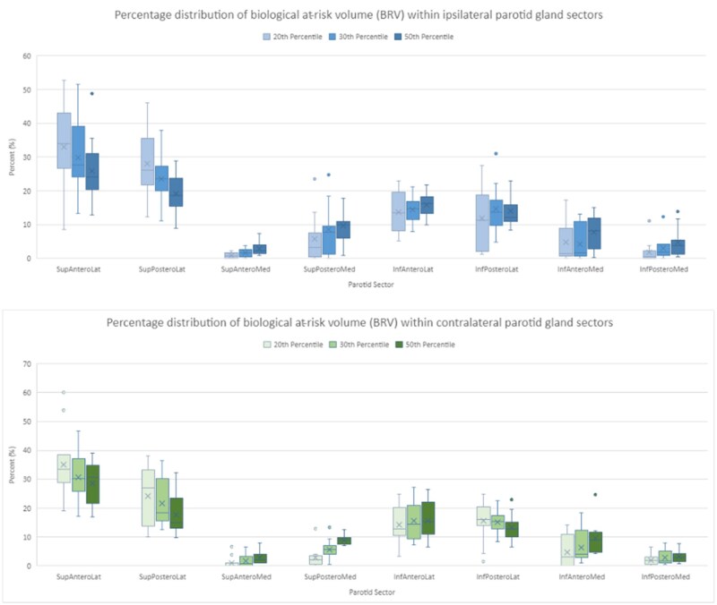
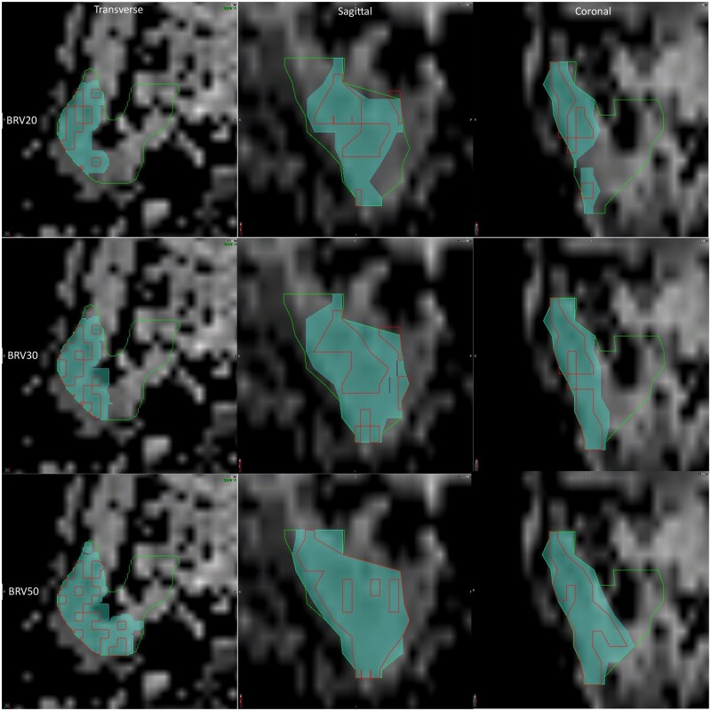
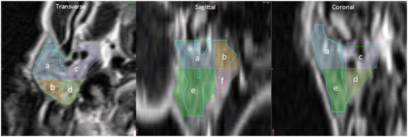
Methods: Image registration between the planning CT and MRI-simulation images was performed and reviewed to ensure accurate translation of ADC data when contouring the PG. Histogram analysis was undertaken using the 20th, 30th, and 50th percentile ADC values of each contoured PG to form the BRV. The whole PG was split into 8 anatomical sectors at a common intersection point to evaluate the distribution of BRV throughout.
Results: The BRV distribution for each percentile was mapped across the whole contoured PG and each anatomical sector contour. The largest distribution was predominantly found in the superolateral sectors.
Conclusions: The 20th and 30th percentile ADC values can be used to form a BRV of the PG. The location of the BRV distribution indicates a potential relationship between ADC thresholds and the functional region of the PG.
Advances in knowledge: The BRV is located in a favourable position within the PG and could be used to further spare this salivary gland during dose optimization. The feasibility of this approach will be explored in a future retrospective dosimetry study.

 求助内容:
求助内容: 应助结果提醒方式:


