{"title":"[牙龈卟啉单胞菌诱导血管内皮细胞铁下垂的作用及机制研究]。","authors":"Q Li, C Lu, J Lin","doi":"10.3760/cma.j.cn112144-20250420-00145","DOIUrl":null,"url":null,"abstract":"<p><p><b>Objective:</b> To investigate whether <i>Porphyromonas gingivalis</i> (Pg) induces ferroptosis in vascular endothelial cells and predict the Hub genes. <b>Methods:</b> Firstly, human umbilical vein endothelial cells (HUVEC) were stimulated with Pg (W83) for 4 h, and transmission electron microscopy was used to observe ferroptosis-related morphological characteristics. Subsequently, RNA was extracted from HUVEC before and after Pg stimulation for transcriptome sequencing (RNA-seq). Enrichment analysis was performed to determine if differentially expressed genes (DEG) associated with ferroptosis. Ferroptosis-related DEG (Fer-DEG) were identified and then underwent gene ontology (GO) functional annotation, Kyoto Encyclopedia of Genes and Genomes (KEGG) pathway enrichment analysis, protein-protein interaction (PPI) network construction, and Hub gene prediction. Next, based on RNA-seq results, HUVEC were stimulated with lipopolysaccharide (LPS) for 24 h. Established ferroptosis markers were detected. The indices and detection methods were as follows: cell viability via cell counting kit-8; reactive oxygen species (ROS) by the DCFH-DA probe; Fe²⁺, lipid peroxides (LPO), malondialdehyde (MDA), and reduced/oxidized glutathione ratio (GSH/GSSG) with commercial kits; mitochondrial membrane potential (MMP) using the JC-1 probe; solute carrier family 7 member 11 (SLC7A11), solute carrier family 3 member 2 (SLC3A2), and glutathione peroxidase 4 (GPX4) expressions by Western blotting (WB) and real-time fluorescence quantitative PCR (RT-qPCR). Finally, RT-qPCR was used to validate the expression of predicted Hub genes in HUVEC after 24 h LPS stimulation, including tumor necrosis factor (TNF) or TNF-α, interleukin (IL)-6, and prostaglandin-endoperoxide synthase 2 (PTGS2). <b>Results:</b> The mitochondria exhibited size reduction and cristae loss in Pg-stimulated HUVEC. DEG of HUVEC between the Pg-infected and control groups were enriched in the pathway of ferroptosis, and from which 56 Fer-DEG were identified. GO analysis showed enrichment in in responses to TNF, LPS, biotic stimulus, etc. and KEGG analysis revealed enrichment in TNF, C-type lectin receptor, and IL-17 signaling pathways, etc. In the 56-gene PPI network, TNF, IL-6, and PTGS2 were predicted as Hub genes, which were significantly associated with ferroptosis-related pathways, including unsaturated fatty acid biosynthesis and ROS metabolic process regulation. Compared to the control group [(100.00±1.44)%], LPS significantly reduced HUVEC viability [(66.77±1.80)%], which could be ameliorated by Fer-1 [(84.50±1.47)%] (<i>P<</i>0.05). The ROS fluorescence intensity in the LPS group (1 523.00±250.70) was significantly higher than in the control (328.20±38.68) or LPS+Fer-1 (753.30±67.11) group (all <i>P<</i>0.05). The Fe²⁺, LPO, and MDA levels in the LPS group [(29.83±4.25) μmol/10<sup>6</sup> cells, (3.58±0.24) μmol/gprot, (5.54±0.33) μmol/gprot, respectively] were significantly higher than both the control group [(7.29±0.79) μmol/10<sup>6</sup> cells, (1.08±0.05) μmol/gprot, (2.06±0.17) μmol/gprot] and the LPS+Fer-1 group [(16.33±1.63) μmol/10<sup>6</sup> cells, (2.01±0.09) μmol/gprot, (3.24±0.26) μmol/gprot]. Furthermore, the GSH/GSSG ratio in the LPS group (2.17±0.08) was considerably lower than both the control group (6.96±0.20) and the LPS+Fer-1 group (4.31±0.81) (all <i>P<</i>0.05). The JC-1 aggregate/monomer fluorescence intensity ratio of the LPS group (0.46±0.07) was markedly lower than the control group (285.60±160.40), while Fer-1 pretreatment (1.53±0.17) obviously mitigated this decrease (all <i>P<</i>0.05). SLC7A11, SLC3A2, and GPX4 protein and mRNA expression levels in the LPS group were dramatically lower than both the control group and the Fer-1+LPS group (<i>P<</i>0.05). The mRNA expression levels of TNF, IL-6, and PTGS2 in the LPS group were strongly upregulated compared to the control group, and the expressions of these three factors in the LPS+Fer-1 group were significantly lower than those in the LPS group (all <i>P<</i>0.05). <b>Conclusions:</b> Pg drives ferroptosis in vascular endothelial cells, with TNF, IL-6, and PTGS2 identified as the potential novel Hub genes in this process.</p>","PeriodicalId":23965,"journal":{"name":"中华口腔医学杂志","volume":"60 9","pages":"1008-1018"},"PeriodicalIF":0.0000,"publicationDate":"2025-09-09","publicationTypes":"Journal Article","fieldsOfStudy":null,"isOpenAccess":false,"openAccessPdf":"","citationCount":"0","resultStr":"{\"title\":\"[Investigation of the role and mechanism of <i>Porphyromonas gingivalis</i> in inducing ferroptosis in vascular endothelial cells].\",\"authors\":\"Q Li, C Lu, J Lin\",\"doi\":\"10.3760/cma.j.cn112144-20250420-00145\",\"DOIUrl\":null,\"url\":null,\"abstract\":\"<p><p><b>Objective:</b> To investigate whether <i>Porphyromonas gingivalis</i> (Pg) induces ferroptosis in vascular endothelial cells and predict the Hub genes. <b>Methods:</b> Firstly, human umbilical vein endothelial cells (HUVEC) were stimulated with Pg (W83) for 4 h, and transmission electron microscopy was used to observe ferroptosis-related morphological characteristics. Subsequently, RNA was extracted from HUVEC before and after Pg stimulation for transcriptome sequencing (RNA-seq). Enrichment analysis was performed to determine if differentially expressed genes (DEG) associated with ferroptosis. Ferroptosis-related DEG (Fer-DEG) were identified and then underwent gene ontology (GO) functional annotation, Kyoto Encyclopedia of Genes and Genomes (KEGG) pathway enrichment analysis, protein-protein interaction (PPI) network construction, and Hub gene prediction. Next, based on RNA-seq results, HUVEC were stimulated with lipopolysaccharide (LPS) for 24 h. Established ferroptosis markers were detected. The indices and detection methods were as follows: cell viability via cell counting kit-8; reactive oxygen species (ROS) by the DCFH-DA probe; Fe²⁺, lipid peroxides (LPO), malondialdehyde (MDA), and reduced/oxidized glutathione ratio (GSH/GSSG) with commercial kits; mitochondrial membrane potential (MMP) using the JC-1 probe; solute carrier family 7 member 11 (SLC7A11), solute carrier family 3 member 2 (SLC3A2), and glutathione peroxidase 4 (GPX4) expressions by Western blotting (WB) and real-time fluorescence quantitative PCR (RT-qPCR). Finally, RT-qPCR was used to validate the expression of predicted Hub genes in HUVEC after 24 h LPS stimulation, including tumor necrosis factor (TNF) or TNF-α, interleukin (IL)-6, and prostaglandin-endoperoxide synthase 2 (PTGS2). <b>Results:</b> The mitochondria exhibited size reduction and cristae loss in Pg-stimulated HUVEC. DEG of HUVEC between the Pg-infected and control groups were enriched in the pathway of ferroptosis, and from which 56 Fer-DEG were identified. GO analysis showed enrichment in in responses to TNF, LPS, biotic stimulus, etc. and KEGG analysis revealed enrichment in TNF, C-type lectin receptor, and IL-17 signaling pathways, etc. In the 56-gene PPI network, TNF, IL-6, and PTGS2 were predicted as Hub genes, which were significantly associated with ferroptosis-related pathways, including unsaturated fatty acid biosynthesis and ROS metabolic process regulation. Compared to the control group [(100.00±1.44)%], LPS significantly reduced HUVEC viability [(66.77±1.80)%], which could be ameliorated by Fer-1 [(84.50±1.47)%] (<i>P<</i>0.05). The ROS fluorescence intensity in the LPS group (1 523.00±250.70) was significantly higher than in the control (328.20±38.68) or LPS+Fer-1 (753.30±67.11) group (all <i>P<</i>0.05). The Fe²⁺, LPO, and MDA levels in the LPS group [(29.83±4.25) μmol/10<sup>6</sup> cells, (3.58±0.24) μmol/gprot, (5.54±0.33) μmol/gprot, respectively] were significantly higher than both the control group [(7.29±0.79) μmol/10<sup>6</sup> cells, (1.08±0.05) μmol/gprot, (2.06±0.17) μmol/gprot] and the LPS+Fer-1 group [(16.33±1.63) μmol/10<sup>6</sup> cells, (2.01±0.09) μmol/gprot, (3.24±0.26) μmol/gprot]. Furthermore, the GSH/GSSG ratio in the LPS group (2.17±0.08) was considerably lower than both the control group (6.96±0.20) and the LPS+Fer-1 group (4.31±0.81) (all <i>P<</i>0.05). The JC-1 aggregate/monomer fluorescence intensity ratio of the LPS group (0.46±0.07) was markedly lower than the control group (285.60±160.40), while Fer-1 pretreatment (1.53±0.17) obviously mitigated this decrease (all <i>P<</i>0.05). SLC7A11, SLC3A2, and GPX4 protein and mRNA expression levels in the LPS group were dramatically lower than both the control group and the Fer-1+LPS group (<i>P<</i>0.05). The mRNA expression levels of TNF, IL-6, and PTGS2 in the LPS group were strongly upregulated compared to the control group, and the expressions of these three factors in the LPS+Fer-1 group were significantly lower than those in the LPS group (all <i>P<</i>0.05). <b>Conclusions:</b> Pg drives ferroptosis in vascular endothelial cells, with TNF, IL-6, and PTGS2 identified as the potential novel Hub genes in this process.</p>\",\"PeriodicalId\":23965,\"journal\":{\"name\":\"中华口腔医学杂志\",\"volume\":\"60 9\",\"pages\":\"1008-1018\"},\"PeriodicalIF\":0.0000,\"publicationDate\":\"2025-09-09\",\"publicationTypes\":\"Journal Article\",\"fieldsOfStudy\":null,\"isOpenAccess\":false,\"openAccessPdf\":\"\",\"citationCount\":\"0\",\"resultStr\":null,\"platform\":\"Semanticscholar\",\"paperid\":null,\"PeriodicalName\":\"中华口腔医学杂志\",\"FirstCategoryId\":\"3\",\"ListUrlMain\":\"https://doi.org/10.3760/cma.j.cn112144-20250420-00145\",\"RegionNum\":0,\"RegionCategory\":null,\"ArticlePicture\":[],\"TitleCN\":null,\"AbstractTextCN\":null,\"PMCID\":null,\"EPubDate\":\"\",\"PubModel\":\"\",\"JCR\":\"Q4\",\"JCRName\":\"Medicine\",\"Score\":null,\"Total\":0}","platform":"Semanticscholar","paperid":null,"PeriodicalName":"中华口腔医学杂志","FirstCategoryId":"3","ListUrlMain":"https://doi.org/10.3760/cma.j.cn112144-20250420-00145","RegionNum":0,"RegionCategory":null,"ArticlePicture":[],"TitleCN":null,"AbstractTextCN":null,"PMCID":null,"EPubDate":"","PubModel":"","JCR":"Q4","JCRName":"Medicine","Score":null,"Total":0}
[Investigation of the role and mechanism of Porphyromonas gingivalis in inducing ferroptosis in vascular endothelial cells].
Objective: To investigate whether Porphyromonas gingivalis (Pg) induces ferroptosis in vascular endothelial cells and predict the Hub genes. Methods: Firstly, human umbilical vein endothelial cells (HUVEC) were stimulated with Pg (W83) for 4 h, and transmission electron microscopy was used to observe ferroptosis-related morphological characteristics. Subsequently, RNA was extracted from HUVEC before and after Pg stimulation for transcriptome sequencing (RNA-seq). Enrichment analysis was performed to determine if differentially expressed genes (DEG) associated with ferroptosis. Ferroptosis-related DEG (Fer-DEG) were identified and then underwent gene ontology (GO) functional annotation, Kyoto Encyclopedia of Genes and Genomes (KEGG) pathway enrichment analysis, protein-protein interaction (PPI) network construction, and Hub gene prediction. Next, based on RNA-seq results, HUVEC were stimulated with lipopolysaccharide (LPS) for 24 h. Established ferroptosis markers were detected. The indices and detection methods were as follows: cell viability via cell counting kit-8; reactive oxygen species (ROS) by the DCFH-DA probe; Fe²⁺, lipid peroxides (LPO), malondialdehyde (MDA), and reduced/oxidized glutathione ratio (GSH/GSSG) with commercial kits; mitochondrial membrane potential (MMP) using the JC-1 probe; solute carrier family 7 member 11 (SLC7A11), solute carrier family 3 member 2 (SLC3A2), and glutathione peroxidase 4 (GPX4) expressions by Western blotting (WB) and real-time fluorescence quantitative PCR (RT-qPCR). Finally, RT-qPCR was used to validate the expression of predicted Hub genes in HUVEC after 24 h LPS stimulation, including tumor necrosis factor (TNF) or TNF-α, interleukin (IL)-6, and prostaglandin-endoperoxide synthase 2 (PTGS2). Results: The mitochondria exhibited size reduction and cristae loss in Pg-stimulated HUVEC. DEG of HUVEC between the Pg-infected and control groups were enriched in the pathway of ferroptosis, and from which 56 Fer-DEG were identified. GO analysis showed enrichment in in responses to TNF, LPS, biotic stimulus, etc. and KEGG analysis revealed enrichment in TNF, C-type lectin receptor, and IL-17 signaling pathways, etc. In the 56-gene PPI network, TNF, IL-6, and PTGS2 were predicted as Hub genes, which were significantly associated with ferroptosis-related pathways, including unsaturated fatty acid biosynthesis and ROS metabolic process regulation. Compared to the control group [(100.00±1.44)%], LPS significantly reduced HUVEC viability [(66.77±1.80)%], which could be ameliorated by Fer-1 [(84.50±1.47)%] (P<0.05). The ROS fluorescence intensity in the LPS group (1 523.00±250.70) was significantly higher than in the control (328.20±38.68) or LPS+Fer-1 (753.30±67.11) group (all P<0.05). The Fe²⁺, LPO, and MDA levels in the LPS group [(29.83±4.25) μmol/106 cells, (3.58±0.24) μmol/gprot, (5.54±0.33) μmol/gprot, respectively] were significantly higher than both the control group [(7.29±0.79) μmol/106 cells, (1.08±0.05) μmol/gprot, (2.06±0.17) μmol/gprot] and the LPS+Fer-1 group [(16.33±1.63) μmol/106 cells, (2.01±0.09) μmol/gprot, (3.24±0.26) μmol/gprot]. Furthermore, the GSH/GSSG ratio in the LPS group (2.17±0.08) was considerably lower than both the control group (6.96±0.20) and the LPS+Fer-1 group (4.31±0.81) (all P<0.05). The JC-1 aggregate/monomer fluorescence intensity ratio of the LPS group (0.46±0.07) was markedly lower than the control group (285.60±160.40), while Fer-1 pretreatment (1.53±0.17) obviously mitigated this decrease (all P<0.05). SLC7A11, SLC3A2, and GPX4 protein and mRNA expression levels in the LPS group were dramatically lower than both the control group and the Fer-1+LPS group (P<0.05). The mRNA expression levels of TNF, IL-6, and PTGS2 in the LPS group were strongly upregulated compared to the control group, and the expressions of these three factors in the LPS+Fer-1 group were significantly lower than those in the LPS group (all P<0.05). Conclusions: Pg drives ferroptosis in vascular endothelial cells, with TNF, IL-6, and PTGS2 identified as the potential novel Hub genes in this process.
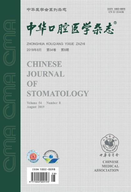
期刊介绍:
Founded in August 1953, Chinese Journal of Stomatology is a monthly academic journal of stomatology published publicly at home and abroad, sponsored by the Chinese Medical Association and co-sponsored by the Chinese Stomatology Association. It mainly reports the leading scientific research results and clinical diagnosis and treatment experience in the field of oral medicine, as well as the basic theoretical research that has a guiding role in oral clinical practice and is closely combined with oral clinical practice.
Chinese Journal of Over the years, Stomatology has been published in Medline, Scopus database, Toxicology Abstracts Database, Chemical Abstracts Database, American Cancer database, Russian Abstracts database, China Core Journal of Science and Technology, Peking University Core Journal, CSCD and other more than 20 important journals at home and abroad Physical medicine database and retrieval system included.