{"title":"基于磁共振成像的放射组学特征预测食管癌术前分期。","authors":"Ri-Hui Yang, Zhi-Ping Lin, Ting Dong, Wei-Xiong Fan, Hao-Dong Qin, Gui-Hua Jiang, Hai-Yang Dai","doi":"10.4329/wjr.v17.i8.110307","DOIUrl":null,"url":null,"abstract":"<p><strong>Background: </strong>Esophageal cancer (EC) is one of the most prevalent malignant gastrointestinal tumors; accurate prediction of EC staging has high significance before treatment.</p><p><strong>Aim: </strong>To explore a rational radiomic approach for predicting preoperative staging of EC based on magnetic resonance imaging (MRI).</p><p><strong>Methods: </strong>This retrospective study included 210 patients with pathologically confirmed EC, randomly divided into a primary cohort (<i>n</i> = 147) and a validation cohort (<i>n</i> = 63) in a ratio of 7:3. All patients underwent a preoperative MRI scan from the neck to the abdomen. High-throughput and quantitative radiomics features were extracted from T2-weighted imaging (T2WI) and gadolinium contrast-enhanced T1-weighted imaging (T1WI)-Gd images. Radiomics signatures were selected using minimal redundancy maximal relevance and the least absolute shrinkage and selection operator. Then a logistic regression model was built to predict the EC stages. The diagnostic performance of the radiomics model for discriminating between stages I-II and III-IV was evaluated using the area under the curve (AUC), sensitivity (SEN), and specificity (SPE).</p><p><strong>Results: </strong>A total of 214 radiomics features were extracted. Following feature dimension reduction, the T1WI and T2WI sequences were retained, and 14 features from the T1WI sequence and 3 features from the T2WI sequence were selected to construct radiomics signatures. The radiomics signature combining T2WI with T1WI-Gd demonstrated superior discrimination of stages in the validation cohort (AUC: 0.851; SEN: 0.697; SPE: 0.793), which outperformed single-sequence models (AUC: 0.779, 0.844; SEN: 0.667, 0.636; SPE: 0.8, 0.8).</p><p><strong>Conclusion: </strong>MRI-based radiomics signatures could identify EC stages before treatment, which could serve as a noninvasive and quantitative approach aiding personalized treatment planning.</p>","PeriodicalId":23819,"journal":{"name":"World journal of radiology","volume":"17 8","pages":"110307"},"PeriodicalIF":1.5000,"publicationDate":"2025-08-28","publicationTypes":"Journal Article","fieldsOfStudy":null,"isOpenAccess":false,"openAccessPdf":"https://www.ncbi.nlm.nih.gov/pmc/articles/PMC12400284/pdf/","citationCount":"0","resultStr":"{\"title\":\"Magnetic resonance imaging-based radiomics signature for predicting preoperative staging of esophageal cancer.\",\"authors\":\"Ri-Hui Yang, Zhi-Ping Lin, Ting Dong, Wei-Xiong Fan, Hao-Dong Qin, Gui-Hua Jiang, Hai-Yang Dai\",\"doi\":\"10.4329/wjr.v17.i8.110307\",\"DOIUrl\":null,\"url\":null,\"abstract\":\"<p><strong>Background: </strong>Esophageal cancer (EC) is one of the most prevalent malignant gastrointestinal tumors; accurate prediction of EC staging has high significance before treatment.</p><p><strong>Aim: </strong>To explore a rational radiomic approach for predicting preoperative staging of EC based on magnetic resonance imaging (MRI).</p><p><strong>Methods: </strong>This retrospective study included 210 patients with pathologically confirmed EC, randomly divided into a primary cohort (<i>n</i> = 147) and a validation cohort (<i>n</i> = 63) in a ratio of 7:3. All patients underwent a preoperative MRI scan from the neck to the abdomen. High-throughput and quantitative radiomics features were extracted from T2-weighted imaging (T2WI) and gadolinium contrast-enhanced T1-weighted imaging (T1WI)-Gd images. Radiomics signatures were selected using minimal redundancy maximal relevance and the least absolute shrinkage and selection operator. Then a logistic regression model was built to predict the EC stages. The diagnostic performance of the radiomics model for discriminating between stages I-II and III-IV was evaluated using the area under the curve (AUC), sensitivity (SEN), and specificity (SPE).</p><p><strong>Results: </strong>A total of 214 radiomics features were extracted. Following feature dimension reduction, the T1WI and T2WI sequences were retained, and 14 features from the T1WI sequence and 3 features from the T2WI sequence were selected to construct radiomics signatures. The radiomics signature combining T2WI with T1WI-Gd demonstrated superior discrimination of stages in the validation cohort (AUC: 0.851; SEN: 0.697; SPE: 0.793), which outperformed single-sequence models (AUC: 0.779, 0.844; SEN: 0.667, 0.636; SPE: 0.8, 0.8).</p><p><strong>Conclusion: </strong>MRI-based radiomics signatures could identify EC stages before treatment, which could serve as a noninvasive and quantitative approach aiding personalized treatment planning.</p>\",\"PeriodicalId\":23819,\"journal\":{\"name\":\"World journal of radiology\",\"volume\":\"17 8\",\"pages\":\"110307\"},\"PeriodicalIF\":1.5000,\"publicationDate\":\"2025-08-28\",\"publicationTypes\":\"Journal Article\",\"fieldsOfStudy\":null,\"isOpenAccess\":false,\"openAccessPdf\":\"https://www.ncbi.nlm.nih.gov/pmc/articles/PMC12400284/pdf/\",\"citationCount\":\"0\",\"resultStr\":null,\"platform\":\"Semanticscholar\",\"paperid\":null,\"PeriodicalName\":\"World journal of radiology\",\"FirstCategoryId\":\"1085\",\"ListUrlMain\":\"https://doi.org/10.4329/wjr.v17.i8.110307\",\"RegionNum\":0,\"RegionCategory\":null,\"ArticlePicture\":[],\"TitleCN\":null,\"AbstractTextCN\":null,\"PMCID\":null,\"EPubDate\":\"\",\"PubModel\":\"\",\"JCR\":\"Q3\",\"JCRName\":\"RADIOLOGY, NUCLEAR MEDICINE & MEDICAL IMAGING\",\"Score\":null,\"Total\":0}","platform":"Semanticscholar","paperid":null,"PeriodicalName":"World journal of radiology","FirstCategoryId":"1085","ListUrlMain":"https://doi.org/10.4329/wjr.v17.i8.110307","RegionNum":0,"RegionCategory":null,"ArticlePicture":[],"TitleCN":null,"AbstractTextCN":null,"PMCID":null,"EPubDate":"","PubModel":"","JCR":"Q3","JCRName":"RADIOLOGY, NUCLEAR MEDICINE & MEDICAL IMAGING","Score":null,"Total":0}
Magnetic resonance imaging-based radiomics signature for predicting preoperative staging of esophageal cancer.
Background: Esophageal cancer (EC) is one of the most prevalent malignant gastrointestinal tumors; accurate prediction of EC staging has high significance before treatment.
Aim: To explore a rational radiomic approach for predicting preoperative staging of EC based on magnetic resonance imaging (MRI).
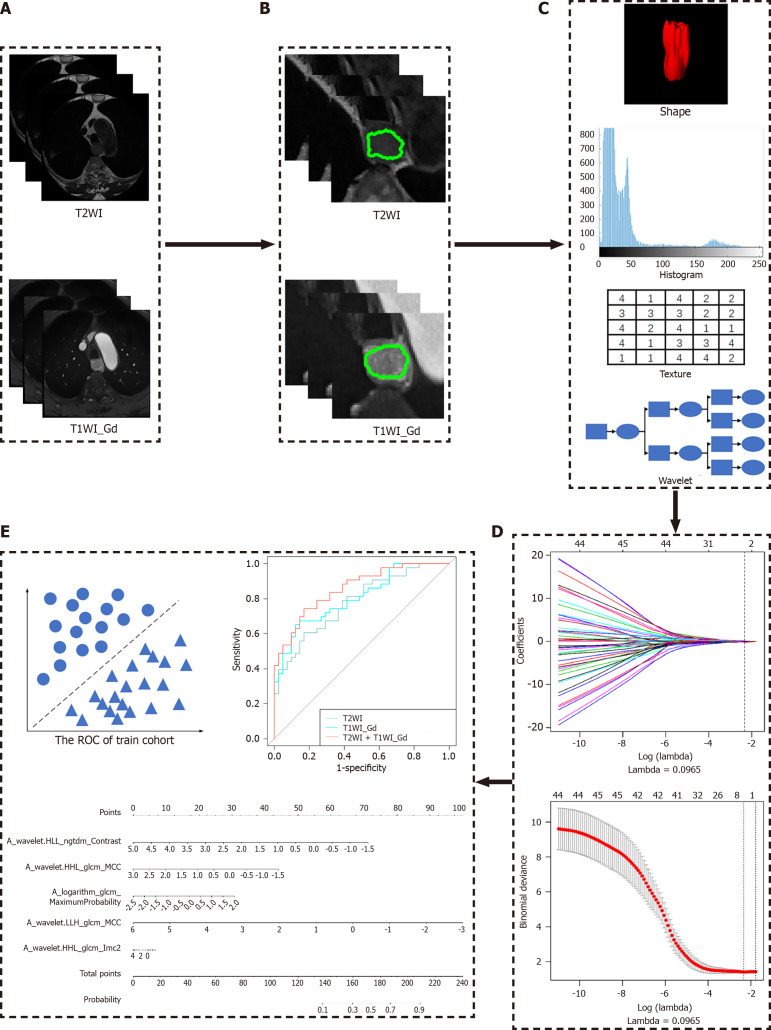
Methods: This retrospective study included 210 patients with pathologically confirmed EC, randomly divided into a primary cohort (n = 147) and a validation cohort (n = 63) in a ratio of 7:3. All patients underwent a preoperative MRI scan from the neck to the abdomen. High-throughput and quantitative radiomics features were extracted from T2-weighted imaging (T2WI) and gadolinium contrast-enhanced T1-weighted imaging (T1WI)-Gd images. Radiomics signatures were selected using minimal redundancy maximal relevance and the least absolute shrinkage and selection operator. Then a logistic regression model was built to predict the EC stages. The diagnostic performance of the radiomics model for discriminating between stages I-II and III-IV was evaluated using the area under the curve (AUC), sensitivity (SEN), and specificity (SPE).
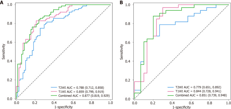
Results: A total of 214 radiomics features were extracted. Following feature dimension reduction, the T1WI and T2WI sequences were retained, and 14 features from the T1WI sequence and 3 features from the T2WI sequence were selected to construct radiomics signatures. The radiomics signature combining T2WI with T1WI-Gd demonstrated superior discrimination of stages in the validation cohort (AUC: 0.851; SEN: 0.697; SPE: 0.793), which outperformed single-sequence models (AUC: 0.779, 0.844; SEN: 0.667, 0.636; SPE: 0.8, 0.8).
Conclusion: MRI-based radiomics signatures could identify EC stages before treatment, which could serve as a noninvasive and quantitative approach aiding personalized treatment planning.

 求助内容:
求助内容: 应助结果提醒方式:


