Carlo Altomare, Rebecca Casati, Giuseppina Pacella, Laura Olivieri, Angelo Tirabasso, Annamaria Altomare, Luca Frasca, Filippo Longo, Pierfilippo Crucitti, Eliodoro Faiella, Bruno Beomonte Zobel, Rosario Francesco Grasso
{"title":"c臂锥束ct引导下的术前微线圈肺磨玻璃结节定位:诊断和手术优势。","authors":"Carlo Altomare, Rebecca Casati, Giuseppina Pacella, Laura Olivieri, Angelo Tirabasso, Annamaria Altomare, Luca Frasca, Filippo Longo, Pierfilippo Crucitti, Eliodoro Faiella, Bruno Beomonte Zobel, Rosario Francesco Grasso","doi":"10.1111/1759-7714.70152","DOIUrl":null,"url":null,"abstract":"<p><strong>Objective: </strong>This study evaluates the effectiveness and safety of C-arm cone beam CT (CBCT)-guided microcoil localization combined with uniportal video-assisted thoracoscopic surgery (VATS) for the management of small, difficult-to-localize ground-glass opacities (GGOs) and sub-solid nodules in the lungs.</p><p><strong>Methods: </strong>We retrospectively analyzed data from 13 patients with single, small, peripheral, non-subpleural GGOs or SSN. All patients underwent successful microcoil localization using CB-CT guidance followed by uniportal VATS resection. A microcoil was positioned partly in the lung parenchyma and partly in the extra-pleural space to assist in intraoperative localization. We evaluated the rate of correct microcoil placement and the technical success of the resection.</p><p><strong>Results: </strong>Microcoil placement was successfully performed in all patients, with an average procedure time of 28.8 ± 10.8 min. The mean nodule size was 9.9 ± 5.4 mm, and 76.9% of the nodules were classified as ground-glass opacities. No intraparenchymal bleeding was observed, and four patients (30.8%) experienced pneumothorax, all of which were self-limited and required no intervention or coil repositioning. The uniVATS resection success rate was 100%.</p><p><strong>Conclusion: </strong>CBCT-guided microcoil localization, with partial placement of the coil in the extra-pleural space, proved to be a highly effective technique for the localization and resection of small pulmonary nodules. The procedure demonstrated high accuracy, minimal complications, reduction of procedural time, and short hospital stays. Intraoperative fluoroscopy was never necessary, with a high reduction in radiation exposure for the patient and the operator. Further studies with larger populations and longer follow-ups are needed to validate these findings.</p>","PeriodicalId":23338,"journal":{"name":"Thoracic Cancer","volume":"16 17","pages":"e70152"},"PeriodicalIF":2.3000,"publicationDate":"2025-09-01","publicationTypes":"Journal Article","fieldsOfStudy":null,"isOpenAccess":false,"openAccessPdf":"https://www.ncbi.nlm.nih.gov/pmc/articles/PMC12408194/pdf/","citationCount":"0","resultStr":"{\"title\":\"C-Arm Cone Beam CT-Guided Preoperative Microcoil Pulmonary Ground Glass Nodule Localization: Diagnostic and Surgical Advantage.\",\"authors\":\"Carlo Altomare, Rebecca Casati, Giuseppina Pacella, Laura Olivieri, Angelo Tirabasso, Annamaria Altomare, Luca Frasca, Filippo Longo, Pierfilippo Crucitti, Eliodoro Faiella, Bruno Beomonte Zobel, Rosario Francesco Grasso\",\"doi\":\"10.1111/1759-7714.70152\",\"DOIUrl\":null,\"url\":null,\"abstract\":\"<p><strong>Objective: </strong>This study evaluates the effectiveness and safety of C-arm cone beam CT (CBCT)-guided microcoil localization combined with uniportal video-assisted thoracoscopic surgery (VATS) for the management of small, difficult-to-localize ground-glass opacities (GGOs) and sub-solid nodules in the lungs.</p><p><strong>Methods: </strong>We retrospectively analyzed data from 13 patients with single, small, peripheral, non-subpleural GGOs or SSN. All patients underwent successful microcoil localization using CB-CT guidance followed by uniportal VATS resection. A microcoil was positioned partly in the lung parenchyma and partly in the extra-pleural space to assist in intraoperative localization. We evaluated the rate of correct microcoil placement and the technical success of the resection.</p><p><strong>Results: </strong>Microcoil placement was successfully performed in all patients, with an average procedure time of 28.8 ± 10.8 min. The mean nodule size was 9.9 ± 5.4 mm, and 76.9% of the nodules were classified as ground-glass opacities. No intraparenchymal bleeding was observed, and four patients (30.8%) experienced pneumothorax, all of which were self-limited and required no intervention or coil repositioning. The uniVATS resection success rate was 100%.</p><p><strong>Conclusion: </strong>CBCT-guided microcoil localization, with partial placement of the coil in the extra-pleural space, proved to be a highly effective technique for the localization and resection of small pulmonary nodules. The procedure demonstrated high accuracy, minimal complications, reduction of procedural time, and short hospital stays. Intraoperative fluoroscopy was never necessary, with a high reduction in radiation exposure for the patient and the operator. Further studies with larger populations and longer follow-ups are needed to validate these findings.</p>\",\"PeriodicalId\":23338,\"journal\":{\"name\":\"Thoracic Cancer\",\"volume\":\"16 17\",\"pages\":\"e70152\"},\"PeriodicalIF\":2.3000,\"publicationDate\":\"2025-09-01\",\"publicationTypes\":\"Journal Article\",\"fieldsOfStudy\":null,\"isOpenAccess\":false,\"openAccessPdf\":\"https://www.ncbi.nlm.nih.gov/pmc/articles/PMC12408194/pdf/\",\"citationCount\":\"0\",\"resultStr\":null,\"platform\":\"Semanticscholar\",\"paperid\":null,\"PeriodicalName\":\"Thoracic Cancer\",\"FirstCategoryId\":\"3\",\"ListUrlMain\":\"https://doi.org/10.1111/1759-7714.70152\",\"RegionNum\":3,\"RegionCategory\":\"医学\",\"ArticlePicture\":[],\"TitleCN\":null,\"AbstractTextCN\":null,\"PMCID\":null,\"EPubDate\":\"\",\"PubModel\":\"\",\"JCR\":\"Q3\",\"JCRName\":\"ONCOLOGY\",\"Score\":null,\"Total\":0}","platform":"Semanticscholar","paperid":null,"PeriodicalName":"Thoracic Cancer","FirstCategoryId":"3","ListUrlMain":"https://doi.org/10.1111/1759-7714.70152","RegionNum":3,"RegionCategory":"医学","ArticlePicture":[],"TitleCN":null,"AbstractTextCN":null,"PMCID":null,"EPubDate":"","PubModel":"","JCR":"Q3","JCRName":"ONCOLOGY","Score":null,"Total":0}
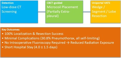
Objective: This study evaluates the effectiveness and safety of C-arm cone beam CT (CBCT)-guided microcoil localization combined with uniportal video-assisted thoracoscopic surgery (VATS) for the management of small, difficult-to-localize ground-glass opacities (GGOs) and sub-solid nodules in the lungs.
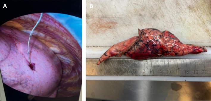
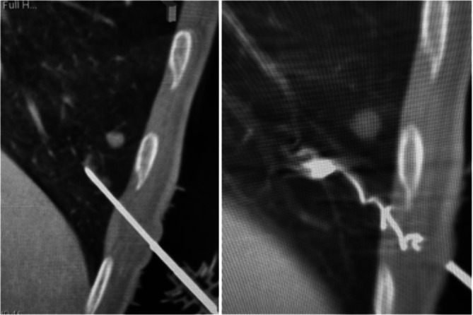
Methods: We retrospectively analyzed data from 13 patients with single, small, peripheral, non-subpleural GGOs or SSN. All patients underwent successful microcoil localization using CB-CT guidance followed by uniportal VATS resection. A microcoil was positioned partly in the lung parenchyma and partly in the extra-pleural space to assist in intraoperative localization. We evaluated the rate of correct microcoil placement and the technical success of the resection.
Results: Microcoil placement was successfully performed in all patients, with an average procedure time of 28.8 ± 10.8 min. The mean nodule size was 9.9 ± 5.4 mm, and 76.9% of the nodules were classified as ground-glass opacities. No intraparenchymal bleeding was observed, and four patients (30.8%) experienced pneumothorax, all of which were self-limited and required no intervention or coil repositioning. The uniVATS resection success rate was 100%.
Conclusion: CBCT-guided microcoil localization, with partial placement of the coil in the extra-pleural space, proved to be a highly effective technique for the localization and resection of small pulmonary nodules. The procedure demonstrated high accuracy, minimal complications, reduction of procedural time, and short hospital stays. Intraoperative fluoroscopy was never necessary, with a high reduction in radiation exposure for the patient and the operator. Further studies with larger populations and longer follow-ups are needed to validate these findings.
期刊介绍:
Thoracic Cancer aims to facilitate international collaboration and exchange of comprehensive and cutting-edge information on basic, translational, and applied clinical research in lung cancer, esophageal cancer, mediastinal cancer, breast cancer and other thoracic malignancies. Prevention, treatment and research relevant to Asia-Pacific is a focus area, but submissions from all regions are welcomed. The editors encourage contributions relevant to prevention, general thoracic surgery, medical oncology, radiology, radiation medicine, pathology, basic cancer research, as well as epidemiological and translational studies in thoracic cancer. Thoracic Cancer is the official publication of the Chinese Society of Lung Cancer, International Chinese Society of Thoracic Surgery and is endorsed by the Korean Association for the Study of Lung Cancer and the Hong Kong Cancer Therapy Society.
The Journal publishes a range of article types including: Editorials, Invited Reviews, Mini Reviews, Original Articles, Clinical Guidelines, Technological Notes, Imaging in thoracic cancer, Meeting Reports, Case Reports, Letters to the Editor, Commentaries, and Brief Reports.
 求助内容:
求助内容: 应助结果提醒方式:


