Ines Lopes Rei, Emilie Paran, Helen Wilson, Mariette Pilot, Alison Catherine Major
{"title":"以脓胸为表现的猫的CT表现及近期预后。","authors":"Ines Lopes Rei, Emilie Paran, Helen Wilson, Mariette Pilot, Alison Catherine Major","doi":"10.1177/1098612X251360637","DOIUrl":null,"url":null,"abstract":"<p><p>ObjectivesThe purpose of this retrospective, descriptive study was to assess CT findings and short-term outcome of cats with pyothorax presented to a referral hospital.MethodsThoracic CT studies of 41 cats with pyothorax comprising pre-contrast lung and soft tissue reconstructions and post-contrast soft tissue reconstructions were blindly reviewed by two European College of Veterinary Diagnostic Imaging (ECVDI) board-certified radiologists and a third-year ECVDI resident, referencing a predetermined list of imaging features. Clinical outcomes, including treatment options, survival to discharge and length of hospitalisation, were recorded.ResultsAll cats (100%) had pleural effusion. Common imaging findings included intrathoracic lymphadenomegaly (85%), pleural thickening (85%), presence of pannus (81%) and evidence of pneumonia (49%). Pulmonary abscessation was suspected in 22% of cats and foreign bodies in 12%. Of the 41 cats, 20 were managed medically, 20 underwent surgery and one had unsuccessful medical treatment followed by surgical intervention. The median duration of hospitalisation was 7 days, and 36 cats survived to discharge.Conclusions and relevanceThis study demonstrates that cats with pyothorax commonly have marked changes on CT that involve multiple thoracic compartments. Our study population highlights that feline pyothorax can have a good short-term outcome when managed either medically or surgically.</p>","PeriodicalId":15851,"journal":{"name":"Journal of Feline Medicine and Surgery","volume":"27 9","pages":"1098612X251360637"},"PeriodicalIF":2.1000,"publicationDate":"2025-09-01","publicationTypes":"Journal Article","fieldsOfStudy":null,"isOpenAccess":false,"openAccessPdf":"https://www.ncbi.nlm.nih.gov/pmc/articles/PMC12411727/pdf/","citationCount":"0","resultStr":"{\"title\":\"CT features and short-term outcome in cats presenting with pyothorax.\",\"authors\":\"Ines Lopes Rei, Emilie Paran, Helen Wilson, Mariette Pilot, Alison Catherine Major\",\"doi\":\"10.1177/1098612X251360637\",\"DOIUrl\":null,\"url\":null,\"abstract\":\"<p><p>ObjectivesThe purpose of this retrospective, descriptive study was to assess CT findings and short-term outcome of cats with pyothorax presented to a referral hospital.MethodsThoracic CT studies of 41 cats with pyothorax comprising pre-contrast lung and soft tissue reconstructions and post-contrast soft tissue reconstructions were blindly reviewed by two European College of Veterinary Diagnostic Imaging (ECVDI) board-certified radiologists and a third-year ECVDI resident, referencing a predetermined list of imaging features. Clinical outcomes, including treatment options, survival to discharge and length of hospitalisation, were recorded.ResultsAll cats (100%) had pleural effusion. Common imaging findings included intrathoracic lymphadenomegaly (85%), pleural thickening (85%), presence of pannus (81%) and evidence of pneumonia (49%). Pulmonary abscessation was suspected in 22% of cats and foreign bodies in 12%. Of the 41 cats, 20 were managed medically, 20 underwent surgery and one had unsuccessful medical treatment followed by surgical intervention. The median duration of hospitalisation was 7 days, and 36 cats survived to discharge.Conclusions and relevanceThis study demonstrates that cats with pyothorax commonly have marked changes on CT that involve multiple thoracic compartments. Our study population highlights that feline pyothorax can have a good short-term outcome when managed either medically or surgically.</p>\",\"PeriodicalId\":15851,\"journal\":{\"name\":\"Journal of Feline Medicine and Surgery\",\"volume\":\"27 9\",\"pages\":\"1098612X251360637\"},\"PeriodicalIF\":2.1000,\"publicationDate\":\"2025-09-01\",\"publicationTypes\":\"Journal Article\",\"fieldsOfStudy\":null,\"isOpenAccess\":false,\"openAccessPdf\":\"https://www.ncbi.nlm.nih.gov/pmc/articles/PMC12411727/pdf/\",\"citationCount\":\"0\",\"resultStr\":null,\"platform\":\"Semanticscholar\",\"paperid\":null,\"PeriodicalName\":\"Journal of Feline Medicine and Surgery\",\"FirstCategoryId\":\"97\",\"ListUrlMain\":\"https://doi.org/10.1177/1098612X251360637\",\"RegionNum\":2,\"RegionCategory\":\"农林科学\",\"ArticlePicture\":[],\"TitleCN\":null,\"AbstractTextCN\":null,\"PMCID\":null,\"EPubDate\":\"2025/9/4 0:00:00\",\"PubModel\":\"Epub\",\"JCR\":\"Q2\",\"JCRName\":\"VETERINARY SCIENCES\",\"Score\":null,\"Total\":0}","platform":"Semanticscholar","paperid":null,"PeriodicalName":"Journal of Feline Medicine and Surgery","FirstCategoryId":"97","ListUrlMain":"https://doi.org/10.1177/1098612X251360637","RegionNum":2,"RegionCategory":"农林科学","ArticlePicture":[],"TitleCN":null,"AbstractTextCN":null,"PMCID":null,"EPubDate":"2025/9/4 0:00:00","PubModel":"Epub","JCR":"Q2","JCRName":"VETERINARY SCIENCES","Score":null,"Total":0}
CT features and short-term outcome in cats presenting with pyothorax.
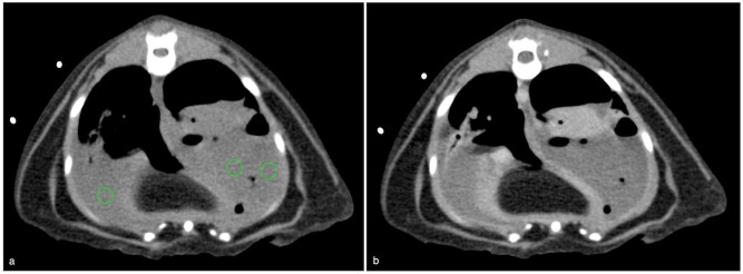
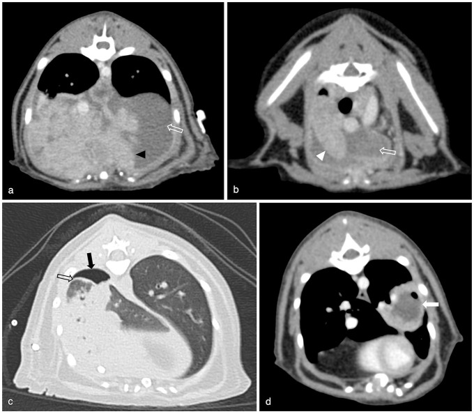
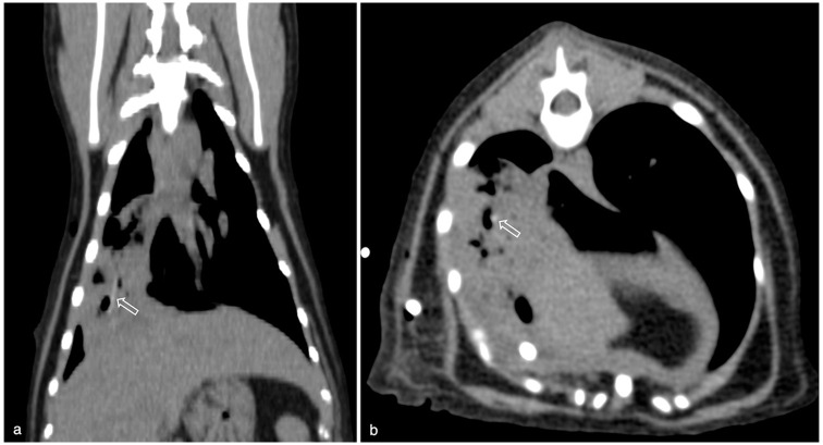
ObjectivesThe purpose of this retrospective, descriptive study was to assess CT findings and short-term outcome of cats with pyothorax presented to a referral hospital.MethodsThoracic CT studies of 41 cats with pyothorax comprising pre-contrast lung and soft tissue reconstructions and post-contrast soft tissue reconstructions were blindly reviewed by two European College of Veterinary Diagnostic Imaging (ECVDI) board-certified radiologists and a third-year ECVDI resident, referencing a predetermined list of imaging features. Clinical outcomes, including treatment options, survival to discharge and length of hospitalisation, were recorded.ResultsAll cats (100%) had pleural effusion. Common imaging findings included intrathoracic lymphadenomegaly (85%), pleural thickening (85%), presence of pannus (81%) and evidence of pneumonia (49%). Pulmonary abscessation was suspected in 22% of cats and foreign bodies in 12%. Of the 41 cats, 20 were managed medically, 20 underwent surgery and one had unsuccessful medical treatment followed by surgical intervention. The median duration of hospitalisation was 7 days, and 36 cats survived to discharge.Conclusions and relevanceThis study demonstrates that cats with pyothorax commonly have marked changes on CT that involve multiple thoracic compartments. Our study population highlights that feline pyothorax can have a good short-term outcome when managed either medically or surgically.
期刊介绍:
JFMS is an international, peer-reviewed journal aimed at both practitioners and researchers with an interest in the clinical veterinary healthcare of domestic cats. The journal is published monthly in two formats: ‘Classic’ editions containing high-quality original papers on all aspects of feline medicine and surgery, including basic research relevant to clinical practice; and dedicated ‘Clinical Practice’ editions primarily containing opinionated review articles providing state-of-the-art information for feline clinicians, along with other relevant articles such as consensus guidelines.
 求助内容:
求助内容: 应助结果提醒方式:


