{"title":"蓝光视网膜损伤BALB/c小鼠模型的建立。","authors":"Arief Wildan, Banundari Rachmawati, Arief Sjamsulaksan Kartasasmita, Fifin Luthfia Rahmi, Maharani, Hermawan Istiadi, Anditta Syifarahmah, Irwan Nurdiansyah, Noviana Fitri Wulandari, Shofia Salsabilah","doi":"10.4103/JAPTR.JAPTR_32_25","DOIUrl":null,"url":null,"abstract":"<p><p>Blue light exposure can damage the retina, resulting in retinal atrophy and significant vision loss. Currently, no efficient animal models can observe retinal damage caused by blue light within a defined timeframe. Creating a BALB/c mouse model for blue light-induced retinal damage is expected to enhance research focused on the prevention and treatment of age-related macular degeneration. This study explores the potential effect of blue light exposure on the BALB/c mice model by analysing apoptosis and retinal degeneration. Anatomical Pathology Laboratory of Diponegoro University and The Integrated Research and Testing Laboratory of Gadjah Mada University. This study design was a posttest-only control group design. Ten five-week-old BALB/c mice were divided into two groups. The exposure group received 10,000 lux of blue light in the special cage for 2 weeks, 3 h daily. Caspase-3 expression was assessed through polymerase chain reaction testing, and retinal thickness was analyzed using hematoxylin and eosin staining. We used the Shapiro-Wilk test to evaluate data normality. Parametric <i>t</i>-tests and nonparametric Mann-Whitney tests were applied to compare groups, with <i>P</i> < 0.05 considered significant. The average whole retinal thickness of the exposed group was 152.812 ± 20.919 µm, while the control group was 214.948 ± 53.284 µm (<i>P</i> = 0.04). The average caspase-3 expression in the exposed group was 19.03 ± 8.57 µm, while the control group was 5.78 ± 2.63 µm (<i>P</i> = 0.011). This approach, utilizing animal models for blue light exposure, can be employed to learn about retinal damage caused by blue light.</p>","PeriodicalId":14877,"journal":{"name":"Journal of Advanced Pharmaceutical Technology & Research","volume":"16 3","pages":"139-143"},"PeriodicalIF":1.4000,"publicationDate":"2025-07-01","publicationTypes":"Journal Article","fieldsOfStudy":null,"isOpenAccess":false,"openAccessPdf":"https://www.ncbi.nlm.nih.gov/pmc/articles/PMC12401520/pdf/","citationCount":"0","resultStr":"{\"title\":\"Development of a BALB/c mice model for blue light retinal damage.\",\"authors\":\"Arief Wildan, Banundari Rachmawati, Arief Sjamsulaksan Kartasasmita, Fifin Luthfia Rahmi, Maharani, Hermawan Istiadi, Anditta Syifarahmah, Irwan Nurdiansyah, Noviana Fitri Wulandari, Shofia Salsabilah\",\"doi\":\"10.4103/JAPTR.JAPTR_32_25\",\"DOIUrl\":null,\"url\":null,\"abstract\":\"<p><p>Blue light exposure can damage the retina, resulting in retinal atrophy and significant vision loss. Currently, no efficient animal models can observe retinal damage caused by blue light within a defined timeframe. Creating a BALB/c mouse model for blue light-induced retinal damage is expected to enhance research focused on the prevention and treatment of age-related macular degeneration. This study explores the potential effect of blue light exposure on the BALB/c mice model by analysing apoptosis and retinal degeneration. Anatomical Pathology Laboratory of Diponegoro University and The Integrated Research and Testing Laboratory of Gadjah Mada University. This study design was a posttest-only control group design. Ten five-week-old BALB/c mice were divided into two groups. The exposure group received 10,000 lux of blue light in the special cage for 2 weeks, 3 h daily. Caspase-3 expression was assessed through polymerase chain reaction testing, and retinal thickness was analyzed using hematoxylin and eosin staining. We used the Shapiro-Wilk test to evaluate data normality. Parametric <i>t</i>-tests and nonparametric Mann-Whitney tests were applied to compare groups, with <i>P</i> < 0.05 considered significant. The average whole retinal thickness of the exposed group was 152.812 ± 20.919 µm, while the control group was 214.948 ± 53.284 µm (<i>P</i> = 0.04). The average caspase-3 expression in the exposed group was 19.03 ± 8.57 µm, while the control group was 5.78 ± 2.63 µm (<i>P</i> = 0.011). This approach, utilizing animal models for blue light exposure, can be employed to learn about retinal damage caused by blue light.</p>\",\"PeriodicalId\":14877,\"journal\":{\"name\":\"Journal of Advanced Pharmaceutical Technology & Research\",\"volume\":\"16 3\",\"pages\":\"139-143\"},\"PeriodicalIF\":1.4000,\"publicationDate\":\"2025-07-01\",\"publicationTypes\":\"Journal Article\",\"fieldsOfStudy\":null,\"isOpenAccess\":false,\"openAccessPdf\":\"https://www.ncbi.nlm.nih.gov/pmc/articles/PMC12401520/pdf/\",\"citationCount\":\"0\",\"resultStr\":null,\"platform\":\"Semanticscholar\",\"paperid\":null,\"PeriodicalName\":\"Journal of Advanced Pharmaceutical Technology & Research\",\"FirstCategoryId\":\"1085\",\"ListUrlMain\":\"https://doi.org/10.4103/JAPTR.JAPTR_32_25\",\"RegionNum\":0,\"RegionCategory\":null,\"ArticlePicture\":[],\"TitleCN\":null,\"AbstractTextCN\":null,\"PMCID\":null,\"EPubDate\":\"2025/8/9 0:00:00\",\"PubModel\":\"Epub\",\"JCR\":\"Q3\",\"JCRName\":\"Pharmacology, Toxicology and Pharmaceutics\",\"Score\":null,\"Total\":0}","platform":"Semanticscholar","paperid":null,"PeriodicalName":"Journal of Advanced Pharmaceutical Technology & Research","FirstCategoryId":"1085","ListUrlMain":"https://doi.org/10.4103/JAPTR.JAPTR_32_25","RegionNum":0,"RegionCategory":null,"ArticlePicture":[],"TitleCN":null,"AbstractTextCN":null,"PMCID":null,"EPubDate":"2025/8/9 0:00:00","PubModel":"Epub","JCR":"Q3","JCRName":"Pharmacology, Toxicology and Pharmaceutics","Score":null,"Total":0}
Development of a BALB/c mice model for blue light retinal damage.
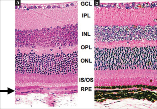
Blue light exposure can damage the retina, resulting in retinal atrophy and significant vision loss. Currently, no efficient animal models can observe retinal damage caused by blue light within a defined timeframe. Creating a BALB/c mouse model for blue light-induced retinal damage is expected to enhance research focused on the prevention and treatment of age-related macular degeneration. This study explores the potential effect of blue light exposure on the BALB/c mice model by analysing apoptosis and retinal degeneration. Anatomical Pathology Laboratory of Diponegoro University and The Integrated Research and Testing Laboratory of Gadjah Mada University. This study design was a posttest-only control group design. Ten five-week-old BALB/c mice were divided into two groups. The exposure group received 10,000 lux of blue light in the special cage for 2 weeks, 3 h daily. Caspase-3 expression was assessed through polymerase chain reaction testing, and retinal thickness was analyzed using hematoxylin and eosin staining. We used the Shapiro-Wilk test to evaluate data normality. Parametric t-tests and nonparametric Mann-Whitney tests were applied to compare groups, with P < 0.05 considered significant. The average whole retinal thickness of the exposed group was 152.812 ± 20.919 µm, while the control group was 214.948 ± 53.284 µm (P = 0.04). The average caspase-3 expression in the exposed group was 19.03 ± 8.57 µm, while the control group was 5.78 ± 2.63 µm (P = 0.011). This approach, utilizing animal models for blue light exposure, can be employed to learn about retinal damage caused by blue light.
期刊介绍:
Journal of Advanced Pharmaceutical Technology & Research (JAPTR) is an Official Publication of Society of Pharmaceutical Education & Research™. It is an international journal published Quarterly. Journal of Advanced Pharmaceutical Technology & Research (JAPTR) is available in online and print version. It is a peer reviewed journal aiming to communicate high quality original research work, reviews, short communications, case report, Ethics Forum, Education Forum and Letter to editor that contribute significantly to further the scientific knowledge related to the field of Pharmacy i.e. Pharmaceutics, Pharmacology, Pharmacognosy, Pharmaceutical Chemistry. Articles with timely interest and newer research concepts will be given more preference.

 求助内容:
求助内容: 应助结果提醒方式:


