Audrey Huang, Jessica Dorilio, Hasit Mehta, Jared M Pisapia
{"title":"在硬膜内脊髓蛛网膜囊肿的手术治疗中,平衡快速场回波序列与术中表现的相关性:说明性病例。","authors":"Audrey Huang, Jessica Dorilio, Hasit Mehta, Jared M Pisapia","doi":"10.3171/CASE25145","DOIUrl":null,"url":null,"abstract":"<p><strong>Background: </strong>Spinal arachnoid cysts can cause myelopathy through spinal cord compression. While MRI is the standard for diagnosis, traditional sequences may not clearly define cyst borders and septations, which are important for guiding surgical intervention. Balanced fast field echo (B-FFE) is an MRI sequence that highlights small arachnoid membranes within and at the borders of CSF spaces.</p><p><strong>Observations: </strong>The authors report the case of a 13-year-old female who presented with progressive lower extremity paresthesias and weakness and urinary incontinence. MRI revealed an intradural cervicothoracic arachnoid cyst (C7-T3) dorsal to the spinal cord. B-FFE was used to identify the upper and lower borders of the intradural arachnoid cyst and its internal septations. These findings corresponded precisely with intraoperative findings and guided fenestration at the cyst's cranial, caudal, and internal septal ends. Postoperatively, the patient's symptoms resolved, and MRI confirmed the resolution of mass effect. At the 10- and 30-month follow-ups, there was no evidence of cyst recurrence clinically or radiographically.</p><p><strong>Lessons: </strong>The authors raise awareness of the clinical utility of B-FFE imaging for intradural spinal arachnoid cysts. Due to its ability to demonstrate cyst borders and internal septations, it offers an alternative to more invasive tests, especially in the pediatric population. https://thejns.org/doi/10.3171/CASE25145.</p>","PeriodicalId":94098,"journal":{"name":"Journal of neurosurgery. Case lessons","volume":"10 9","pages":""},"PeriodicalIF":0.0000,"publicationDate":"2025-09-01","publicationTypes":"Journal Article","fieldsOfStudy":null,"isOpenAccess":false,"openAccessPdf":"https://www.ncbi.nlm.nih.gov/pmc/articles/PMC12400851/pdf/","citationCount":"0","resultStr":"{\"title\":\"Correlation between balanced fast field echo sequence and intraoperative findings in the surgical treatment of an intradural spinal arachnoid cyst: illustrative case.\",\"authors\":\"Audrey Huang, Jessica Dorilio, Hasit Mehta, Jared M Pisapia\",\"doi\":\"10.3171/CASE25145\",\"DOIUrl\":null,\"url\":null,\"abstract\":\"<p><strong>Background: </strong>Spinal arachnoid cysts can cause myelopathy through spinal cord compression. While MRI is the standard for diagnosis, traditional sequences may not clearly define cyst borders and septations, which are important for guiding surgical intervention. Balanced fast field echo (B-FFE) is an MRI sequence that highlights small arachnoid membranes within and at the borders of CSF spaces.</p><p><strong>Observations: </strong>The authors report the case of a 13-year-old female who presented with progressive lower extremity paresthesias and weakness and urinary incontinence. MRI revealed an intradural cervicothoracic arachnoid cyst (C7-T3) dorsal to the spinal cord. B-FFE was used to identify the upper and lower borders of the intradural arachnoid cyst and its internal septations. These findings corresponded precisely with intraoperative findings and guided fenestration at the cyst's cranial, caudal, and internal septal ends. Postoperatively, the patient's symptoms resolved, and MRI confirmed the resolution of mass effect. At the 10- and 30-month follow-ups, there was no evidence of cyst recurrence clinically or radiographically.</p><p><strong>Lessons: </strong>The authors raise awareness of the clinical utility of B-FFE imaging for intradural spinal arachnoid cysts. Due to its ability to demonstrate cyst borders and internal septations, it offers an alternative to more invasive tests, especially in the pediatric population. https://thejns.org/doi/10.3171/CASE25145.</p>\",\"PeriodicalId\":94098,\"journal\":{\"name\":\"Journal of neurosurgery. Case lessons\",\"volume\":\"10 9\",\"pages\":\"\"},\"PeriodicalIF\":0.0000,\"publicationDate\":\"2025-09-01\",\"publicationTypes\":\"Journal Article\",\"fieldsOfStudy\":null,\"isOpenAccess\":false,\"openAccessPdf\":\"https://www.ncbi.nlm.nih.gov/pmc/articles/PMC12400851/pdf/\",\"citationCount\":\"0\",\"resultStr\":null,\"platform\":\"Semanticscholar\",\"paperid\":null,\"PeriodicalName\":\"Journal of neurosurgery. Case lessons\",\"FirstCategoryId\":\"1085\",\"ListUrlMain\":\"https://doi.org/10.3171/CASE25145\",\"RegionNum\":0,\"RegionCategory\":null,\"ArticlePicture\":[],\"TitleCN\":null,\"AbstractTextCN\":null,\"PMCID\":null,\"EPubDate\":\"\",\"PubModel\":\"\",\"JCR\":\"\",\"JCRName\":\"\",\"Score\":null,\"Total\":0}","platform":"Semanticscholar","paperid":null,"PeriodicalName":"Journal of neurosurgery. Case lessons","FirstCategoryId":"1085","ListUrlMain":"https://doi.org/10.3171/CASE25145","RegionNum":0,"RegionCategory":null,"ArticlePicture":[],"TitleCN":null,"AbstractTextCN":null,"PMCID":null,"EPubDate":"","PubModel":"","JCR":"","JCRName":"","Score":null,"Total":0}
Correlation between balanced fast field echo sequence and intraoperative findings in the surgical treatment of an intradural spinal arachnoid cyst: illustrative case.
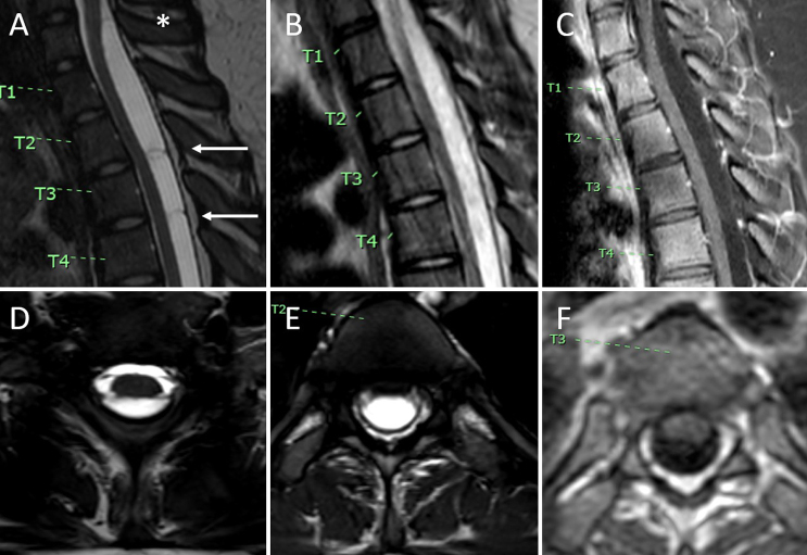
Background: Spinal arachnoid cysts can cause myelopathy through spinal cord compression. While MRI is the standard for diagnosis, traditional sequences may not clearly define cyst borders and septations, which are important for guiding surgical intervention. Balanced fast field echo (B-FFE) is an MRI sequence that highlights small arachnoid membranes within and at the borders of CSF spaces.
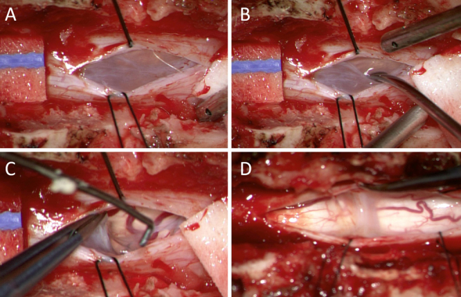
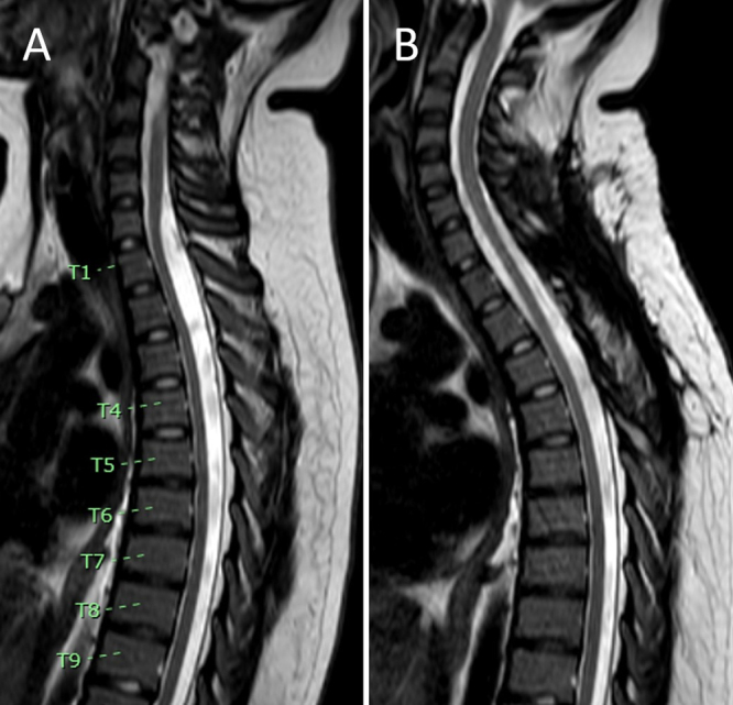
Observations: The authors report the case of a 13-year-old female who presented with progressive lower extremity paresthesias and weakness and urinary incontinence. MRI revealed an intradural cervicothoracic arachnoid cyst (C7-T3) dorsal to the spinal cord. B-FFE was used to identify the upper and lower borders of the intradural arachnoid cyst and its internal septations. These findings corresponded precisely with intraoperative findings and guided fenestration at the cyst's cranial, caudal, and internal septal ends. Postoperatively, the patient's symptoms resolved, and MRI confirmed the resolution of mass effect. At the 10- and 30-month follow-ups, there was no evidence of cyst recurrence clinically or radiographically.
Lessons: The authors raise awareness of the clinical utility of B-FFE imaging for intradural spinal arachnoid cysts. Due to its ability to demonstrate cyst borders and internal septations, it offers an alternative to more invasive tests, especially in the pediatric population. https://thejns.org/doi/10.3171/CASE25145.

 求助内容:
求助内容: 应助结果提醒方式:


