Maciej Laskowski, Bartłomiej Błaszczyk, Marcin Setlak, Adam Rudnik, Ewa Warmuz-Uhma, Jan Herzyk
{"title":"前庭神经鞘瘤治疗后胼胝体脾脏的回飞征:1例报告及文献复习。","authors":"Maciej Laskowski, Bartłomiej Błaszczyk, Marcin Setlak, Adam Rudnik, Ewa Warmuz-Uhma, Jan Herzyk","doi":"10.3390/reports8030136","DOIUrl":null,"url":null,"abstract":"<p><p><b>Background and Clinical Significance</b>: The term \"boomerang sign\" refers to a boomerang-shaped area of cytotoxic edema in the splenium of the corpus callosum. It is seen as hyperintense lesions on T2-weighted images, FLAIR and DWI in MRI. No specific pathomechanism leading to these changes in the splenium have been yet found; however, authors have listed a variety of potential causes. <b>Case Presentation</b>: The case presents a 38-year-old male patient after left cerebellopontine angle tumor resection with an abnormal, increased signal intensity within the corpus callosum (boomerang sign) in FLAIR MRI sequence. In the case of our patient, unlike the patients described in the literature, the changes in the commissure persist. <b>Conclusions</b>: These lesions could be caused by several factors such as the development of cerebellar edema and subarachnoid bleeding or hypertonic salt usage while in the intensive care unit.</p>","PeriodicalId":74664,"journal":{"name":"Reports (MDPI)","volume":"8 3","pages":""},"PeriodicalIF":0.8000,"publicationDate":"2025-08-04","publicationTypes":"Journal Article","fieldsOfStudy":null,"isOpenAccess":false,"openAccessPdf":"https://www.ncbi.nlm.nih.gov/pmc/articles/PMC12372113/pdf/","citationCount":"0","resultStr":"{\"title\":\"Boomerang Sign in the Splenium of the Corpus Callosum After Vestibullar Schwannoma Treatment: Case Report and Review of the Literature.\",\"authors\":\"Maciej Laskowski, Bartłomiej Błaszczyk, Marcin Setlak, Adam Rudnik, Ewa Warmuz-Uhma, Jan Herzyk\",\"doi\":\"10.3390/reports8030136\",\"DOIUrl\":null,\"url\":null,\"abstract\":\"<p><p><b>Background and Clinical Significance</b>: The term \\\"boomerang sign\\\" refers to a boomerang-shaped area of cytotoxic edema in the splenium of the corpus callosum. It is seen as hyperintense lesions on T2-weighted images, FLAIR and DWI in MRI. No specific pathomechanism leading to these changes in the splenium have been yet found; however, authors have listed a variety of potential causes. <b>Case Presentation</b>: The case presents a 38-year-old male patient after left cerebellopontine angle tumor resection with an abnormal, increased signal intensity within the corpus callosum (boomerang sign) in FLAIR MRI sequence. In the case of our patient, unlike the patients described in the literature, the changes in the commissure persist. <b>Conclusions</b>: These lesions could be caused by several factors such as the development of cerebellar edema and subarachnoid bleeding or hypertonic salt usage while in the intensive care unit.</p>\",\"PeriodicalId\":74664,\"journal\":{\"name\":\"Reports (MDPI)\",\"volume\":\"8 3\",\"pages\":\"\"},\"PeriodicalIF\":0.8000,\"publicationDate\":\"2025-08-04\",\"publicationTypes\":\"Journal Article\",\"fieldsOfStudy\":null,\"isOpenAccess\":false,\"openAccessPdf\":\"https://www.ncbi.nlm.nih.gov/pmc/articles/PMC12372113/pdf/\",\"citationCount\":\"0\",\"resultStr\":null,\"platform\":\"Semanticscholar\",\"paperid\":null,\"PeriodicalName\":\"Reports (MDPI)\",\"FirstCategoryId\":\"1085\",\"ListUrlMain\":\"https://doi.org/10.3390/reports8030136\",\"RegionNum\":0,\"RegionCategory\":null,\"ArticlePicture\":[],\"TitleCN\":null,\"AbstractTextCN\":null,\"PMCID\":null,\"EPubDate\":\"\",\"PubModel\":\"\",\"JCR\":\"Q3\",\"JCRName\":\"MEDICINE, GENERAL & INTERNAL\",\"Score\":null,\"Total\":0}","platform":"Semanticscholar","paperid":null,"PeriodicalName":"Reports (MDPI)","FirstCategoryId":"1085","ListUrlMain":"https://doi.org/10.3390/reports8030136","RegionNum":0,"RegionCategory":null,"ArticlePicture":[],"TitleCN":null,"AbstractTextCN":null,"PMCID":null,"EPubDate":"","PubModel":"","JCR":"Q3","JCRName":"MEDICINE, GENERAL & INTERNAL","Score":null,"Total":0}
Boomerang Sign in the Splenium of the Corpus Callosum After Vestibullar Schwannoma Treatment: Case Report and Review of the Literature.
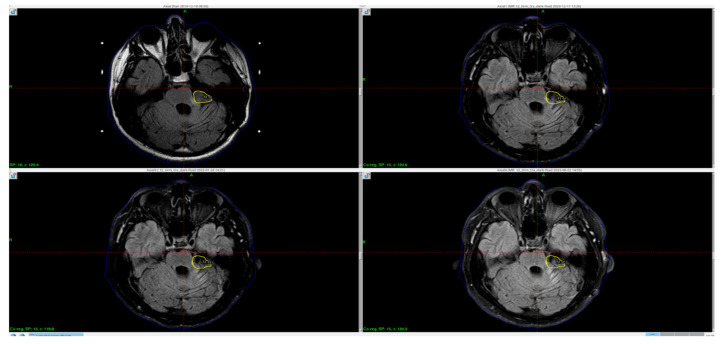
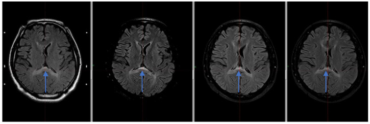
Background and Clinical Significance: The term "boomerang sign" refers to a boomerang-shaped area of cytotoxic edema in the splenium of the corpus callosum. It is seen as hyperintense lesions on T2-weighted images, FLAIR and DWI in MRI. No specific pathomechanism leading to these changes in the splenium have been yet found; however, authors have listed a variety of potential causes. Case Presentation: The case presents a 38-year-old male patient after left cerebellopontine angle tumor resection with an abnormal, increased signal intensity within the corpus callosum (boomerang sign) in FLAIR MRI sequence. In the case of our patient, unlike the patients described in the literature, the changes in the commissure persist. Conclusions: These lesions could be caused by several factors such as the development of cerebellar edema and subarachnoid bleeding or hypertonic salt usage while in the intensive care unit.

 求助内容:
求助内容: 应助结果提醒方式:


