{"title":"蓝痣和蓝痣中产生的黑色素瘤:一个比较的组织病理学病例系列。","authors":"Hristo Popov, Pavel Pavlov, George S Stoyanov","doi":"10.3390/reports8030131","DOIUrl":null,"url":null,"abstract":"<p><p><b>Background and Clinical Significance:</b> Blue nevi are a dubious pigmented lesion. While somewhat common throughout the population, they are significantly less common than other melanocytic neoplasms, and both their morphology and development bring them closer to true hamartomas than neoplasms. An exceedingly rare occurrence is the development of melanoma from a preexisting blue nevus. This nosological unit, defined as melanoma arising in a blue nevus, also known as malignant blue nevus, blue naevus-like melanoma, melanoma ex-blue naevus, and melanoma mimicking cellular blue naevus, is required to either originate from an area of previously excised blue nevus or have a blue nevus remnant adjacent to it. Due to the spindle cell morphology of melanoma arising in blue nevus, the terminology is often misused by some authors to include spindle cell melanomas, which exhibit a distinct pathogenesis and, although morphologically similar, have differing molecular profiles as well. <b>Case presentations:</b> The following manuscript discusses comparative morphological features in a case series of blue nevi and melanoma arising in blue nevi. <b>Discussion:</b> Blue nevi present with unique morphological features, with melanomas originating from them having a unique molecular pathology profile, which significantly differs from other cutaneous melanomas and is closer to that of uveal melanomas.</p>","PeriodicalId":74664,"journal":{"name":"Reports (MDPI)","volume":"8 3","pages":""},"PeriodicalIF":0.8000,"publicationDate":"2025-08-01","publicationTypes":"Journal Article","fieldsOfStudy":null,"isOpenAccess":false,"openAccessPdf":"https://www.ncbi.nlm.nih.gov/pmc/articles/PMC12371955/pdf/","citationCount":"0","resultStr":"{\"title\":\"Blue Nevi and Melanoma Arising in Blue Nevus: A Comparative Histopathological Case Series.\",\"authors\":\"Hristo Popov, Pavel Pavlov, George S Stoyanov\",\"doi\":\"10.3390/reports8030131\",\"DOIUrl\":null,\"url\":null,\"abstract\":\"<p><p><b>Background and Clinical Significance:</b> Blue nevi are a dubious pigmented lesion. While somewhat common throughout the population, they are significantly less common than other melanocytic neoplasms, and both their morphology and development bring them closer to true hamartomas than neoplasms. An exceedingly rare occurrence is the development of melanoma from a preexisting blue nevus. This nosological unit, defined as melanoma arising in a blue nevus, also known as malignant blue nevus, blue naevus-like melanoma, melanoma ex-blue naevus, and melanoma mimicking cellular blue naevus, is required to either originate from an area of previously excised blue nevus or have a blue nevus remnant adjacent to it. Due to the spindle cell morphology of melanoma arising in blue nevus, the terminology is often misused by some authors to include spindle cell melanomas, which exhibit a distinct pathogenesis and, although morphologically similar, have differing molecular profiles as well. <b>Case presentations:</b> The following manuscript discusses comparative morphological features in a case series of blue nevi and melanoma arising in blue nevi. <b>Discussion:</b> Blue nevi present with unique morphological features, with melanomas originating from them having a unique molecular pathology profile, which significantly differs from other cutaneous melanomas and is closer to that of uveal melanomas.</p>\",\"PeriodicalId\":74664,\"journal\":{\"name\":\"Reports (MDPI)\",\"volume\":\"8 3\",\"pages\":\"\"},\"PeriodicalIF\":0.8000,\"publicationDate\":\"2025-08-01\",\"publicationTypes\":\"Journal Article\",\"fieldsOfStudy\":null,\"isOpenAccess\":false,\"openAccessPdf\":\"https://www.ncbi.nlm.nih.gov/pmc/articles/PMC12371955/pdf/\",\"citationCount\":\"0\",\"resultStr\":null,\"platform\":\"Semanticscholar\",\"paperid\":null,\"PeriodicalName\":\"Reports (MDPI)\",\"FirstCategoryId\":\"1085\",\"ListUrlMain\":\"https://doi.org/10.3390/reports8030131\",\"RegionNum\":0,\"RegionCategory\":null,\"ArticlePicture\":[],\"TitleCN\":null,\"AbstractTextCN\":null,\"PMCID\":null,\"EPubDate\":\"\",\"PubModel\":\"\",\"JCR\":\"Q3\",\"JCRName\":\"MEDICINE, GENERAL & INTERNAL\",\"Score\":null,\"Total\":0}","platform":"Semanticscholar","paperid":null,"PeriodicalName":"Reports (MDPI)","FirstCategoryId":"1085","ListUrlMain":"https://doi.org/10.3390/reports8030131","RegionNum":0,"RegionCategory":null,"ArticlePicture":[],"TitleCN":null,"AbstractTextCN":null,"PMCID":null,"EPubDate":"","PubModel":"","JCR":"Q3","JCRName":"MEDICINE, GENERAL & INTERNAL","Score":null,"Total":0}
Blue Nevi and Melanoma Arising in Blue Nevus: A Comparative Histopathological Case Series.
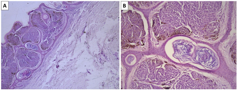
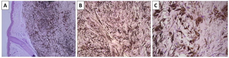
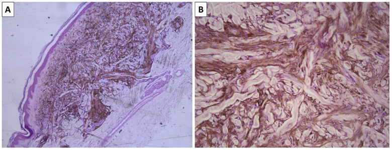
Background and Clinical Significance: Blue nevi are a dubious pigmented lesion. While somewhat common throughout the population, they are significantly less common than other melanocytic neoplasms, and both their morphology and development bring them closer to true hamartomas than neoplasms. An exceedingly rare occurrence is the development of melanoma from a preexisting blue nevus. This nosological unit, defined as melanoma arising in a blue nevus, also known as malignant blue nevus, blue naevus-like melanoma, melanoma ex-blue naevus, and melanoma mimicking cellular blue naevus, is required to either originate from an area of previously excised blue nevus or have a blue nevus remnant adjacent to it. Due to the spindle cell morphology of melanoma arising in blue nevus, the terminology is often misused by some authors to include spindle cell melanomas, which exhibit a distinct pathogenesis and, although morphologically similar, have differing molecular profiles as well. Case presentations: The following manuscript discusses comparative morphological features in a case series of blue nevi and melanoma arising in blue nevi. Discussion: Blue nevi present with unique morphological features, with melanomas originating from them having a unique molecular pathology profile, which significantly differs from other cutaneous melanomas and is closer to that of uveal melanomas.

 求助内容:
求助内容: 应助结果提醒方式:


