George S Stoyanov, Ivaylo Balabanov, Svetoslava Zhivkova, Hristo Popov
{"title":"黑色素成毛细胞瘤:一罕见的色素变异型成毛细胞瘤的组织病理学病例报告。","authors":"George S Stoyanov, Ivaylo Balabanov, Svetoslava Zhivkova, Hristo Popov","doi":"10.3390/reports8030130","DOIUrl":null,"url":null,"abstract":"<p><p><b>Background and clinical significance:</b> Trichoblastomas are rare, mixed epithelial tumors with a mesenchymal component and hair follicle differentiation. <b>Case presentation:</b> Herein, we present a case report of a 51-year-old female patient presenting to the obstetrics and gynecology department with complaints of edema and erythema of the right Bartholin gland, and a lesion measuring 2 cm on the right lateral edge of the mons pubis, towards the inguinal fold. Marsupialization of the Bartholin gland was performed, as well as an incision into the pubo-inguinal lesion, which the patient depicted as grossly resembling an ingrown hair. Upon incision into the pubic-inguinal lesion, it was dark brown in color and spontaneously popped out of the subcutis, without an attempt at enucleation. Histology and subsequent immunohistochemistry of the lesion showed a blue basaloid tumor with an extensive pigment component located deep in the dermis that was sharply demarcated from the surrounding tissues. <b>Conclusion:</b> Immunohistochemistry was diffusely and strongly positive for epithelial markers; melanocytic markers were positive only in dendritic melanocytes dispersed within the tumors, and the proliferative index was low. As such, the tumor was identified as melanotrichoblastoma.</p>","PeriodicalId":74664,"journal":{"name":"Reports (MDPI)","volume":"8 3","pages":""},"PeriodicalIF":0.8000,"publicationDate":"2025-08-01","publicationTypes":"Journal Article","fieldsOfStudy":null,"isOpenAccess":false,"openAccessPdf":"https://www.ncbi.nlm.nih.gov/pmc/articles/PMC12372001/pdf/","citationCount":"0","resultStr":"{\"title\":\"Melanotrichoblastoma: A Histopathological Case Report of a Rare Pigmented Variant of Trichoblastoma.\",\"authors\":\"George S Stoyanov, Ivaylo Balabanov, Svetoslava Zhivkova, Hristo Popov\",\"doi\":\"10.3390/reports8030130\",\"DOIUrl\":null,\"url\":null,\"abstract\":\"<p><p><b>Background and clinical significance:</b> Trichoblastomas are rare, mixed epithelial tumors with a mesenchymal component and hair follicle differentiation. <b>Case presentation:</b> Herein, we present a case report of a 51-year-old female patient presenting to the obstetrics and gynecology department with complaints of edema and erythema of the right Bartholin gland, and a lesion measuring 2 cm on the right lateral edge of the mons pubis, towards the inguinal fold. Marsupialization of the Bartholin gland was performed, as well as an incision into the pubo-inguinal lesion, which the patient depicted as grossly resembling an ingrown hair. Upon incision into the pubic-inguinal lesion, it was dark brown in color and spontaneously popped out of the subcutis, without an attempt at enucleation. Histology and subsequent immunohistochemistry of the lesion showed a blue basaloid tumor with an extensive pigment component located deep in the dermis that was sharply demarcated from the surrounding tissues. <b>Conclusion:</b> Immunohistochemistry was diffusely and strongly positive for epithelial markers; melanocytic markers were positive only in dendritic melanocytes dispersed within the tumors, and the proliferative index was low. As such, the tumor was identified as melanotrichoblastoma.</p>\",\"PeriodicalId\":74664,\"journal\":{\"name\":\"Reports (MDPI)\",\"volume\":\"8 3\",\"pages\":\"\"},\"PeriodicalIF\":0.8000,\"publicationDate\":\"2025-08-01\",\"publicationTypes\":\"Journal Article\",\"fieldsOfStudy\":null,\"isOpenAccess\":false,\"openAccessPdf\":\"https://www.ncbi.nlm.nih.gov/pmc/articles/PMC12372001/pdf/\",\"citationCount\":\"0\",\"resultStr\":null,\"platform\":\"Semanticscholar\",\"paperid\":null,\"PeriodicalName\":\"Reports (MDPI)\",\"FirstCategoryId\":\"1085\",\"ListUrlMain\":\"https://doi.org/10.3390/reports8030130\",\"RegionNum\":0,\"RegionCategory\":null,\"ArticlePicture\":[],\"TitleCN\":null,\"AbstractTextCN\":null,\"PMCID\":null,\"EPubDate\":\"\",\"PubModel\":\"\",\"JCR\":\"Q3\",\"JCRName\":\"MEDICINE, GENERAL & INTERNAL\",\"Score\":null,\"Total\":0}","platform":"Semanticscholar","paperid":null,"PeriodicalName":"Reports (MDPI)","FirstCategoryId":"1085","ListUrlMain":"https://doi.org/10.3390/reports8030130","RegionNum":0,"RegionCategory":null,"ArticlePicture":[],"TitleCN":null,"AbstractTextCN":null,"PMCID":null,"EPubDate":"","PubModel":"","JCR":"Q3","JCRName":"MEDICINE, GENERAL & INTERNAL","Score":null,"Total":0}
Melanotrichoblastoma: A Histopathological Case Report of a Rare Pigmented Variant of Trichoblastoma.
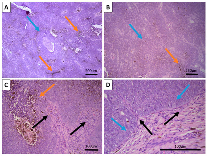
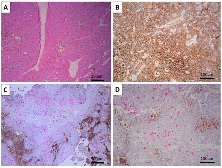
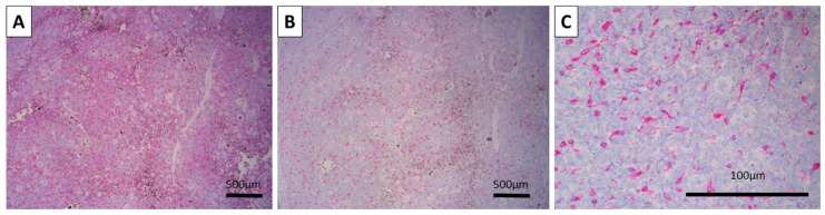
Background and clinical significance: Trichoblastomas are rare, mixed epithelial tumors with a mesenchymal component and hair follicle differentiation. Case presentation: Herein, we present a case report of a 51-year-old female patient presenting to the obstetrics and gynecology department with complaints of edema and erythema of the right Bartholin gland, and a lesion measuring 2 cm on the right lateral edge of the mons pubis, towards the inguinal fold. Marsupialization of the Bartholin gland was performed, as well as an incision into the pubo-inguinal lesion, which the patient depicted as grossly resembling an ingrown hair. Upon incision into the pubic-inguinal lesion, it was dark brown in color and spontaneously popped out of the subcutis, without an attempt at enucleation. Histology and subsequent immunohistochemistry of the lesion showed a blue basaloid tumor with an extensive pigment component located deep in the dermis that was sharply demarcated from the surrounding tissues. Conclusion: Immunohistochemistry was diffusely and strongly positive for epithelial markers; melanocytic markers were positive only in dendritic melanocytes dispersed within the tumors, and the proliferative index was low. As such, the tumor was identified as melanotrichoblastoma.

 求助内容:
求助内容: 应助结果提醒方式:


