{"title":"广泛胆脂瘤损害整个同侧颅底:通过多通道手术技术切除。","authors":"Lyubomir Rangachev, Julian Rangachev, Tzvetomir Marinov, Sylvia Skelina, Todor M Popov","doi":"10.3390/reports8030148","DOIUrl":null,"url":null,"abstract":"<p><p><b>Background and Clinical Significance</b>: Petrous bone cholesteatoma is a rare and complex condition that poses significant challenges in terms of its diagnosis and treatment. This benign yet locally aggressive lesion can cause extensive destruction of the surrounding structures, potentially leading to serious complications. <b>Case Presentation</b>: We present a case of extensive petrous bone cholesteatoma involving nearly the entire skull base. High-resolution CT and MRI were used to assess the extent of the lesion and its relationship with critical neurovascular structures. The cholesteatoma extended from the petrous apex to the clivus, involving the internal auditory canal and Meckel's cave, encasing the internal carotid artery, and compressing the brainstem. The surgical strategy included combined endoscopic transsphenoidal and transclival techniques with a retrolabyrinthine approach. The endoscopic component provided access to the anterior and central skull base regions, whereas the retrolabyrinthine approach allowed us to gain access to the posterior petrous area. Careful dissection was performed to separate the cholesteatoma from the internal carotid artery and the brainstem. Neuromonitoring was performed throughout the procedure to ensure cranial nerve integrity. This combined approach enabled gross total resection, and postoperative imaging confirmed successful tumor removal. The patient's recovery was uneventful, and no new neurological deficits were observed. <b>Conclusions</b>: The successful management of this complex case demonstrates the efficacy and safety of combining endoscopic surgical approaches for extensive skull base cholesteatomas. This multi-corridor approach allows for maximal tumor resection while also minimizing the risks to critical neurovascular structures.</p>","PeriodicalId":74664,"journal":{"name":"Reports (MDPI)","volume":"8 3","pages":""},"PeriodicalIF":0.8000,"publicationDate":"2025-08-18","publicationTypes":"Journal Article","fieldsOfStudy":null,"isOpenAccess":false,"openAccessPdf":"https://www.ncbi.nlm.nih.gov/pmc/articles/PMC12371988/pdf/","citationCount":"0","resultStr":"{\"title\":\"Extensive Cholesteatoma Compromising the Entire Ipsilateral Skull Base: Excision Through a Multi-Corridor Surgical Technique.\",\"authors\":\"Lyubomir Rangachev, Julian Rangachev, Tzvetomir Marinov, Sylvia Skelina, Todor M Popov\",\"doi\":\"10.3390/reports8030148\",\"DOIUrl\":null,\"url\":null,\"abstract\":\"<p><p><b>Background and Clinical Significance</b>: Petrous bone cholesteatoma is a rare and complex condition that poses significant challenges in terms of its diagnosis and treatment. This benign yet locally aggressive lesion can cause extensive destruction of the surrounding structures, potentially leading to serious complications. <b>Case Presentation</b>: We present a case of extensive petrous bone cholesteatoma involving nearly the entire skull base. High-resolution CT and MRI were used to assess the extent of the lesion and its relationship with critical neurovascular structures. The cholesteatoma extended from the petrous apex to the clivus, involving the internal auditory canal and Meckel's cave, encasing the internal carotid artery, and compressing the brainstem. The surgical strategy included combined endoscopic transsphenoidal and transclival techniques with a retrolabyrinthine approach. The endoscopic component provided access to the anterior and central skull base regions, whereas the retrolabyrinthine approach allowed us to gain access to the posterior petrous area. Careful dissection was performed to separate the cholesteatoma from the internal carotid artery and the brainstem. Neuromonitoring was performed throughout the procedure to ensure cranial nerve integrity. This combined approach enabled gross total resection, and postoperative imaging confirmed successful tumor removal. The patient's recovery was uneventful, and no new neurological deficits were observed. <b>Conclusions</b>: The successful management of this complex case demonstrates the efficacy and safety of combining endoscopic surgical approaches for extensive skull base cholesteatomas. This multi-corridor approach allows for maximal tumor resection while also minimizing the risks to critical neurovascular structures.</p>\",\"PeriodicalId\":74664,\"journal\":{\"name\":\"Reports (MDPI)\",\"volume\":\"8 3\",\"pages\":\"\"},\"PeriodicalIF\":0.8000,\"publicationDate\":\"2025-08-18\",\"publicationTypes\":\"Journal Article\",\"fieldsOfStudy\":null,\"isOpenAccess\":false,\"openAccessPdf\":\"https://www.ncbi.nlm.nih.gov/pmc/articles/PMC12371988/pdf/\",\"citationCount\":\"0\",\"resultStr\":null,\"platform\":\"Semanticscholar\",\"paperid\":null,\"PeriodicalName\":\"Reports (MDPI)\",\"FirstCategoryId\":\"1085\",\"ListUrlMain\":\"https://doi.org/10.3390/reports8030148\",\"RegionNum\":0,\"RegionCategory\":null,\"ArticlePicture\":[],\"TitleCN\":null,\"AbstractTextCN\":null,\"PMCID\":null,\"EPubDate\":\"\",\"PubModel\":\"\",\"JCR\":\"Q3\",\"JCRName\":\"MEDICINE, GENERAL & INTERNAL\",\"Score\":null,\"Total\":0}","platform":"Semanticscholar","paperid":null,"PeriodicalName":"Reports (MDPI)","FirstCategoryId":"1085","ListUrlMain":"https://doi.org/10.3390/reports8030148","RegionNum":0,"RegionCategory":null,"ArticlePicture":[],"TitleCN":null,"AbstractTextCN":null,"PMCID":null,"EPubDate":"","PubModel":"","JCR":"Q3","JCRName":"MEDICINE, GENERAL & INTERNAL","Score":null,"Total":0}
Extensive Cholesteatoma Compromising the Entire Ipsilateral Skull Base: Excision Through a Multi-Corridor Surgical Technique.
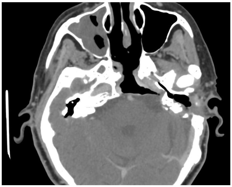
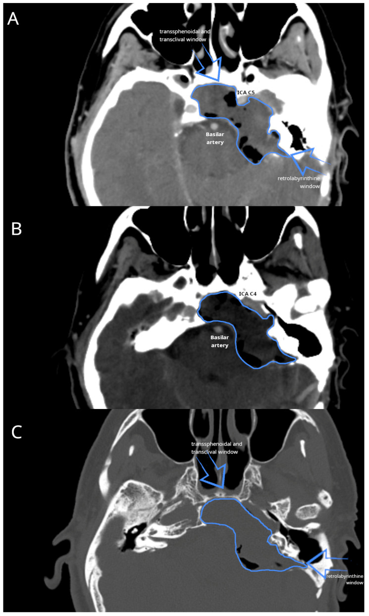
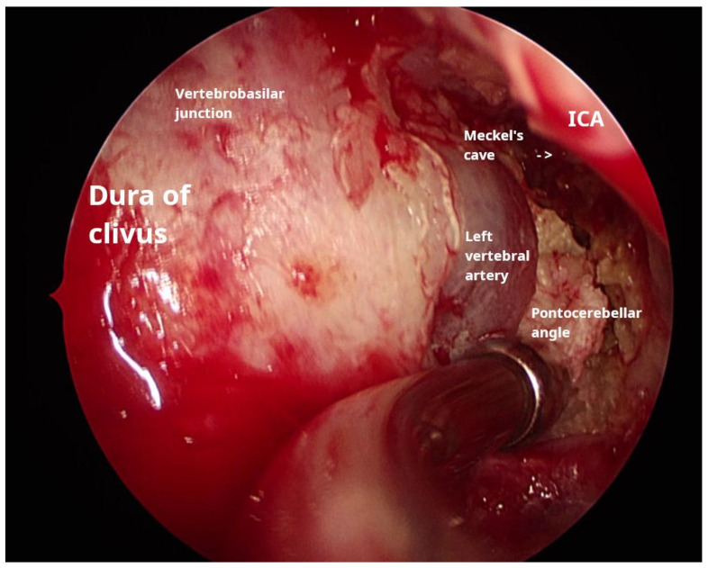
Background and Clinical Significance: Petrous bone cholesteatoma is a rare and complex condition that poses significant challenges in terms of its diagnosis and treatment. This benign yet locally aggressive lesion can cause extensive destruction of the surrounding structures, potentially leading to serious complications. Case Presentation: We present a case of extensive petrous bone cholesteatoma involving nearly the entire skull base. High-resolution CT and MRI were used to assess the extent of the lesion and its relationship with critical neurovascular structures. The cholesteatoma extended from the petrous apex to the clivus, involving the internal auditory canal and Meckel's cave, encasing the internal carotid artery, and compressing the brainstem. The surgical strategy included combined endoscopic transsphenoidal and transclival techniques with a retrolabyrinthine approach. The endoscopic component provided access to the anterior and central skull base regions, whereas the retrolabyrinthine approach allowed us to gain access to the posterior petrous area. Careful dissection was performed to separate the cholesteatoma from the internal carotid artery and the brainstem. Neuromonitoring was performed throughout the procedure to ensure cranial nerve integrity. This combined approach enabled gross total resection, and postoperative imaging confirmed successful tumor removal. The patient's recovery was uneventful, and no new neurological deficits were observed. Conclusions: The successful management of this complex case demonstrates the efficacy and safety of combining endoscopic surgical approaches for extensive skull base cholesteatomas. This multi-corridor approach allows for maximal tumor resection while also minimizing the risks to critical neurovascular structures.

 求助内容:
求助内容: 应助结果提醒方式:


