Mohammad Mehdizadeh, Ali Lotfi, Saede Atarbashi-Moghadam, Parsa Eftekhari Moghadam
{"title":"下颌转移性乳腺癌:罕见病例报告与诊断的挑战。","authors":"Mohammad Mehdizadeh, Ali Lotfi, Saede Atarbashi-Moghadam, Parsa Eftekhari Moghadam","doi":"10.30476/dentjods.2025.103764.2475","DOIUrl":null,"url":null,"abstract":"<p><p>Jawbone metastatic lesions are a diagnostic challenge because of their rarity and variable clinical, radiographic, and histopathologic characteristics. This paper presents a 57-year-old female with a chief complaint of lower face swelling. Cone beam computed tomography (CBCT) showed a multilocular radiolucency with right angle septa in the left mandibular area with cortical destruction. She had a history of right breast cancer about six years ago. Histopathologic examination revealed sheets of malignant small round cells. Immunohistochemistry (IHC) was only positive for cytokeratin (CK) and GATA3. CA15-3 tumor marker was higher than the normal range. Based on the aforementioned data, the diagnosis of metastatic breast carcinoma was performed. The whole-body and computed tomography (CT) scan showed just involvement in the left mandibular area. The radiographic appearance of metastatic lesions might be misleading, and microscopic sections might be poorly differentiated, therefore, a precise past medical history, IHC staining, and tumor markers are valuable issues in diagnosing oral cavity metastasis.</p>","PeriodicalId":73702,"journal":{"name":"Journal of dentistry (Shiraz, Iran)","volume":"26 3","pages":"284-287"},"PeriodicalIF":0.0000,"publicationDate":"2025-09-01","publicationTypes":"Journal Article","fieldsOfStudy":null,"isOpenAccess":false,"openAccessPdf":"https://www.ncbi.nlm.nih.gov/pmc/articles/PMC12394743/pdf/","citationCount":"0","resultStr":"{\"title\":\"Mandibular Metastatic Breast Cancer: A Rare Case Report with Diagnostic Challenge.\",\"authors\":\"Mohammad Mehdizadeh, Ali Lotfi, Saede Atarbashi-Moghadam, Parsa Eftekhari Moghadam\",\"doi\":\"10.30476/dentjods.2025.103764.2475\",\"DOIUrl\":null,\"url\":null,\"abstract\":\"<p><p>Jawbone metastatic lesions are a diagnostic challenge because of their rarity and variable clinical, radiographic, and histopathologic characteristics. This paper presents a 57-year-old female with a chief complaint of lower face swelling. Cone beam computed tomography (CBCT) showed a multilocular radiolucency with right angle septa in the left mandibular area with cortical destruction. She had a history of right breast cancer about six years ago. Histopathologic examination revealed sheets of malignant small round cells. Immunohistochemistry (IHC) was only positive for cytokeratin (CK) and GATA3. CA15-3 tumor marker was higher than the normal range. Based on the aforementioned data, the diagnosis of metastatic breast carcinoma was performed. The whole-body and computed tomography (CT) scan showed just involvement in the left mandibular area. The radiographic appearance of metastatic lesions might be misleading, and microscopic sections might be poorly differentiated, therefore, a precise past medical history, IHC staining, and tumor markers are valuable issues in diagnosing oral cavity metastasis.</p>\",\"PeriodicalId\":73702,\"journal\":{\"name\":\"Journal of dentistry (Shiraz, Iran)\",\"volume\":\"26 3\",\"pages\":\"284-287\"},\"PeriodicalIF\":0.0000,\"publicationDate\":\"2025-09-01\",\"publicationTypes\":\"Journal Article\",\"fieldsOfStudy\":null,\"isOpenAccess\":false,\"openAccessPdf\":\"https://www.ncbi.nlm.nih.gov/pmc/articles/PMC12394743/pdf/\",\"citationCount\":\"0\",\"resultStr\":null,\"platform\":\"Semanticscholar\",\"paperid\":null,\"PeriodicalName\":\"Journal of dentistry (Shiraz, Iran)\",\"FirstCategoryId\":\"1085\",\"ListUrlMain\":\"https://doi.org/10.30476/dentjods.2025.103764.2475\",\"RegionNum\":0,\"RegionCategory\":null,\"ArticlePicture\":[],\"TitleCN\":null,\"AbstractTextCN\":null,\"PMCID\":null,\"EPubDate\":\"\",\"PubModel\":\"\",\"JCR\":\"\",\"JCRName\":\"\",\"Score\":null,\"Total\":0}","platform":"Semanticscholar","paperid":null,"PeriodicalName":"Journal of dentistry (Shiraz, Iran)","FirstCategoryId":"1085","ListUrlMain":"https://doi.org/10.30476/dentjods.2025.103764.2475","RegionNum":0,"RegionCategory":null,"ArticlePicture":[],"TitleCN":null,"AbstractTextCN":null,"PMCID":null,"EPubDate":"","PubModel":"","JCR":"","JCRName":"","Score":null,"Total":0}
Mandibular Metastatic Breast Cancer: A Rare Case Report with Diagnostic Challenge.
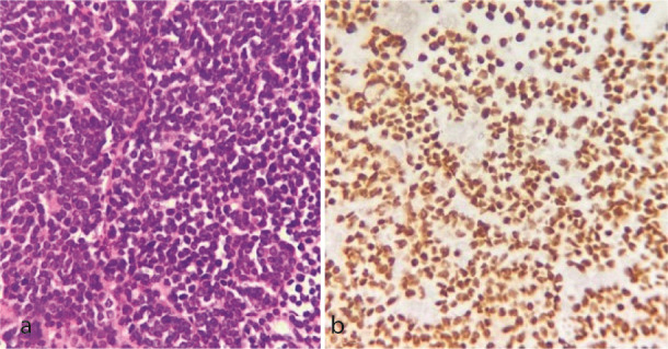
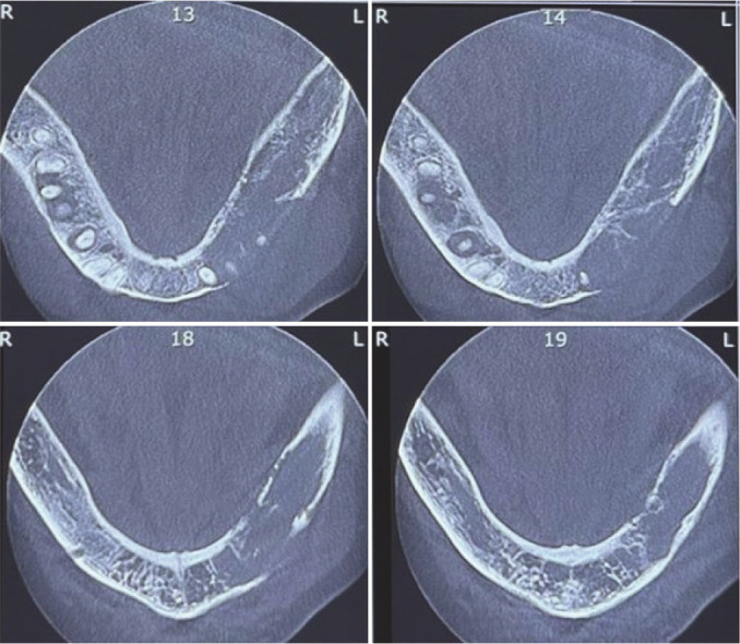
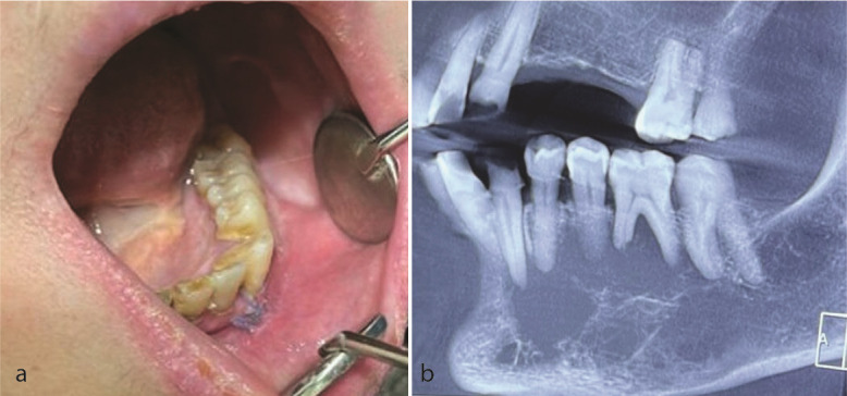
Jawbone metastatic lesions are a diagnostic challenge because of their rarity and variable clinical, radiographic, and histopathologic characteristics. This paper presents a 57-year-old female with a chief complaint of lower face swelling. Cone beam computed tomography (CBCT) showed a multilocular radiolucency with right angle septa in the left mandibular area with cortical destruction. She had a history of right breast cancer about six years ago. Histopathologic examination revealed sheets of malignant small round cells. Immunohistochemistry (IHC) was only positive for cytokeratin (CK) and GATA3. CA15-3 tumor marker was higher than the normal range. Based on the aforementioned data, the diagnosis of metastatic breast carcinoma was performed. The whole-body and computed tomography (CT) scan showed just involvement in the left mandibular area. The radiographic appearance of metastatic lesions might be misleading, and microscopic sections might be poorly differentiated, therefore, a precise past medical history, IHC staining, and tumor markers are valuable issues in diagnosing oral cavity metastasis.

 求助内容:
求助内容: 应助结果提醒方式:


