{"title":"未成熟恒切牙复合牙根骨折后牙根继续形成:1例2年随访报告。","authors":"Maryam Enteghad, Safoora Sahebi, Saleh Hadian","doi":"10.30476/dentjods.2024.104235.2512","DOIUrl":null,"url":null,"abstract":"<p><p>While traumatic injuries in the young permanent dentition are frequent, root fractures are relatively rare, particularly in immature teeth. This study reports the case of a 7-year-old boy who fell off a bicycle. Radiographic examination showed an immature right upper central incisor with fractures in the middle and along the root in an oblique and horizontal direction. Furthermore, there was an extrusion of the coronal segment from its original position. At the first appointment, the right central incisor was repositioned, and a semi-rigid splint was applied for four weeks. The patient was examined periodically for the following two years. After two months, the injured tooth was asymptomatic, with a reduction in probing depth from 8 mm to 2 mm along the tooth surface and a physiologic mobility. Although the injured tooth responded to the electric pulp test after nine months, it had no response to the cold test even after two years. The injured tooth showed continued root maturation of both coronal and apical fragments, although metamorphosis calcification and root canal narrowing were observed in conjunction with mild yellow crown discoloration. This report highlights the ability of Hertwig's epithelial root sheath and immature pulp to continue root development in fractured immature teeth.</p>","PeriodicalId":73702,"journal":{"name":"Journal of dentistry (Shiraz, Iran)","volume":"26 3","pages":"288-292"},"PeriodicalIF":0.0000,"publicationDate":"2025-09-01","publicationTypes":"Journal Article","fieldsOfStudy":null,"isOpenAccess":false,"openAccessPdf":"https://www.ncbi.nlm.nih.gov/pmc/articles/PMC12394745/pdf/","citationCount":"0","resultStr":"{\"title\":\"Continued Root Formation after a Compound root Fracture in an Immature Permanent Incisor: A Case Report with a 2-year follow-up.\",\"authors\":\"Maryam Enteghad, Safoora Sahebi, Saleh Hadian\",\"doi\":\"10.30476/dentjods.2024.104235.2512\",\"DOIUrl\":null,\"url\":null,\"abstract\":\"<p><p>While traumatic injuries in the young permanent dentition are frequent, root fractures are relatively rare, particularly in immature teeth. This study reports the case of a 7-year-old boy who fell off a bicycle. Radiographic examination showed an immature right upper central incisor with fractures in the middle and along the root in an oblique and horizontal direction. Furthermore, there was an extrusion of the coronal segment from its original position. At the first appointment, the right central incisor was repositioned, and a semi-rigid splint was applied for four weeks. The patient was examined periodically for the following two years. After two months, the injured tooth was asymptomatic, with a reduction in probing depth from 8 mm to 2 mm along the tooth surface and a physiologic mobility. Although the injured tooth responded to the electric pulp test after nine months, it had no response to the cold test even after two years. The injured tooth showed continued root maturation of both coronal and apical fragments, although metamorphosis calcification and root canal narrowing were observed in conjunction with mild yellow crown discoloration. This report highlights the ability of Hertwig's epithelial root sheath and immature pulp to continue root development in fractured immature teeth.</p>\",\"PeriodicalId\":73702,\"journal\":{\"name\":\"Journal of dentistry (Shiraz, Iran)\",\"volume\":\"26 3\",\"pages\":\"288-292\"},\"PeriodicalIF\":0.0000,\"publicationDate\":\"2025-09-01\",\"publicationTypes\":\"Journal Article\",\"fieldsOfStudy\":null,\"isOpenAccess\":false,\"openAccessPdf\":\"https://www.ncbi.nlm.nih.gov/pmc/articles/PMC12394745/pdf/\",\"citationCount\":\"0\",\"resultStr\":null,\"platform\":\"Semanticscholar\",\"paperid\":null,\"PeriodicalName\":\"Journal of dentistry (Shiraz, Iran)\",\"FirstCategoryId\":\"1085\",\"ListUrlMain\":\"https://doi.org/10.30476/dentjods.2024.104235.2512\",\"RegionNum\":0,\"RegionCategory\":null,\"ArticlePicture\":[],\"TitleCN\":null,\"AbstractTextCN\":null,\"PMCID\":null,\"EPubDate\":\"\",\"PubModel\":\"\",\"JCR\":\"\",\"JCRName\":\"\",\"Score\":null,\"Total\":0}","platform":"Semanticscholar","paperid":null,"PeriodicalName":"Journal of dentistry (Shiraz, Iran)","FirstCategoryId":"1085","ListUrlMain":"https://doi.org/10.30476/dentjods.2024.104235.2512","RegionNum":0,"RegionCategory":null,"ArticlePicture":[],"TitleCN":null,"AbstractTextCN":null,"PMCID":null,"EPubDate":"","PubModel":"","JCR":"","JCRName":"","Score":null,"Total":0}
Continued Root Formation after a Compound root Fracture in an Immature Permanent Incisor: A Case Report with a 2-year follow-up.
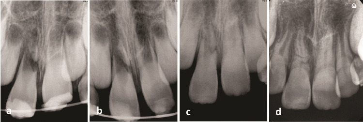
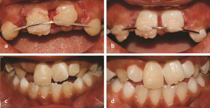
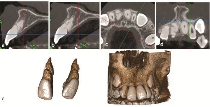
While traumatic injuries in the young permanent dentition are frequent, root fractures are relatively rare, particularly in immature teeth. This study reports the case of a 7-year-old boy who fell off a bicycle. Radiographic examination showed an immature right upper central incisor with fractures in the middle and along the root in an oblique and horizontal direction. Furthermore, there was an extrusion of the coronal segment from its original position. At the first appointment, the right central incisor was repositioned, and a semi-rigid splint was applied for four weeks. The patient was examined periodically for the following two years. After two months, the injured tooth was asymptomatic, with a reduction in probing depth from 8 mm to 2 mm along the tooth surface and a physiologic mobility. Although the injured tooth responded to the electric pulp test after nine months, it had no response to the cold test even after two years. The injured tooth showed continued root maturation of both coronal and apical fragments, although metamorphosis calcification and root canal narrowing were observed in conjunction with mild yellow crown discoloration. This report highlights the ability of Hertwig's epithelial root sheath and immature pulp to continue root development in fractured immature teeth.

 求助内容:
求助内容: 应助结果提醒方式:


