Emma Fransson, Maria Evertsson, Tyra Lundberg, Tebin Hawez, Gustav Andersson, Christina Granéli, Magnus Cinthio, Tobias Erlöv, Pernilla Stenström
{"title":"巨结肠病中区分腺瘤病的组织解剖学特征:一种诊断算法。","authors":"Emma Fransson, Maria Evertsson, Tyra Lundberg, Tebin Hawez, Gustav Andersson, Christina Granéli, Magnus Cinthio, Tobias Erlöv, Pernilla Stenström","doi":"10.3390/diseases13080264","DOIUrl":null,"url":null,"abstract":"<p><strong>Background/objectives: </strong>Intraoperative frozen biopsies are essential during surgery for Hirschsprung's disease (HD). However, this method has several limitations with the need for a faster and real-time diagnostic alternative. For this, consistent histoanatomical and morphometric differences between aganglionic and ganglionic bowel must be established. The primary objective was to compare dimensions of bowel wall layers between aganglionic and ganglionic segments histopathologically in resected rectosigmoid specimens from children with HD. Secondary objectives were to design a diagnostic algorithm to distinguish aganglionosis from ganglionosis and assess whether full bowel wall thickness correlates with patient weight and age.</p><p><strong>Methods: </strong>Each histoanatomic bowel wall layer-mucosa, submucosa, and muscularis propria's layers-was delineated manually on histopathological images. Mean thicknesses were calculated automatically using an in-house image analysis software. Paired parametric tests compared measurements in aganglionic and ganglionic segments.</p><p><strong>Results: </strong>Resected specimens from 30 children with HD were included. Compared to aganglionic bowel, ganglionic bowel showed a thicker muscularis interna (mean 0.666 mm versus 0.461 mm, CI -0.257-(-0.153), <i>p</i> < 0.001), and a higher muscularis interna/muscularis externa ratio (2.047 mm versus 1.287 mm, CI -0.954-(-0.565), <i>p</i> < 0.001). An algorithm based on these features achieved 100% accuracy in distinguishing aganglionosis from ganglionosis. No significant difference in full bowel wall thickness was found between aganglionic and ganglionic segments, nor any correlation with patient weight or age.</p><p><strong>Conclusions: </strong>Histoanatomic layer thickness differs between aganglionic and ganglionic bowel, forming the basis of a diagnostic algorithm. Full bowel wall thickness was independent of patient weight and age.</p>","PeriodicalId":72832,"journal":{"name":"Diseases (Basel, Switzerland)","volume":"13 8","pages":""},"PeriodicalIF":3.0000,"publicationDate":"2025-08-16","publicationTypes":"Journal Article","fieldsOfStudy":null,"isOpenAccess":false,"openAccessPdf":"https://www.ncbi.nlm.nih.gov/pmc/articles/PMC12385927/pdf/","citationCount":"0","resultStr":"{\"title\":\"Histoanatomic Features Distinguishing Aganglionosis in Hirschsprung's Disease: Toward a Diagnostic Algorithm.\",\"authors\":\"Emma Fransson, Maria Evertsson, Tyra Lundberg, Tebin Hawez, Gustav Andersson, Christina Granéli, Magnus Cinthio, Tobias Erlöv, Pernilla Stenström\",\"doi\":\"10.3390/diseases13080264\",\"DOIUrl\":null,\"url\":null,\"abstract\":\"<p><strong>Background/objectives: </strong>Intraoperative frozen biopsies are essential during surgery for Hirschsprung's disease (HD). However, this method has several limitations with the need for a faster and real-time diagnostic alternative. For this, consistent histoanatomical and morphometric differences between aganglionic and ganglionic bowel must be established. The primary objective was to compare dimensions of bowel wall layers between aganglionic and ganglionic segments histopathologically in resected rectosigmoid specimens from children with HD. Secondary objectives were to design a diagnostic algorithm to distinguish aganglionosis from ganglionosis and assess whether full bowel wall thickness correlates with patient weight and age.</p><p><strong>Methods: </strong>Each histoanatomic bowel wall layer-mucosa, submucosa, and muscularis propria's layers-was delineated manually on histopathological images. Mean thicknesses were calculated automatically using an in-house image analysis software. Paired parametric tests compared measurements in aganglionic and ganglionic segments.</p><p><strong>Results: </strong>Resected specimens from 30 children with HD were included. Compared to aganglionic bowel, ganglionic bowel showed a thicker muscularis interna (mean 0.666 mm versus 0.461 mm, CI -0.257-(-0.153), <i>p</i> < 0.001), and a higher muscularis interna/muscularis externa ratio (2.047 mm versus 1.287 mm, CI -0.954-(-0.565), <i>p</i> < 0.001). An algorithm based on these features achieved 100% accuracy in distinguishing aganglionosis from ganglionosis. No significant difference in full bowel wall thickness was found between aganglionic and ganglionic segments, nor any correlation with patient weight or age.</p><p><strong>Conclusions: </strong>Histoanatomic layer thickness differs between aganglionic and ganglionic bowel, forming the basis of a diagnostic algorithm. Full bowel wall thickness was independent of patient weight and age.</p>\",\"PeriodicalId\":72832,\"journal\":{\"name\":\"Diseases (Basel, Switzerland)\",\"volume\":\"13 8\",\"pages\":\"\"},\"PeriodicalIF\":3.0000,\"publicationDate\":\"2025-08-16\",\"publicationTypes\":\"Journal Article\",\"fieldsOfStudy\":null,\"isOpenAccess\":false,\"openAccessPdf\":\"https://www.ncbi.nlm.nih.gov/pmc/articles/PMC12385927/pdf/\",\"citationCount\":\"0\",\"resultStr\":null,\"platform\":\"Semanticscholar\",\"paperid\":null,\"PeriodicalName\":\"Diseases (Basel, Switzerland)\",\"FirstCategoryId\":\"1085\",\"ListUrlMain\":\"https://doi.org/10.3390/diseases13080264\",\"RegionNum\":0,\"RegionCategory\":null,\"ArticlePicture\":[],\"TitleCN\":null,\"AbstractTextCN\":null,\"PMCID\":null,\"EPubDate\":\"\",\"PubModel\":\"\",\"JCR\":\"Q2\",\"JCRName\":\"MEDICINE, RESEARCH & EXPERIMENTAL\",\"Score\":null,\"Total\":0}","platform":"Semanticscholar","paperid":null,"PeriodicalName":"Diseases (Basel, Switzerland)","FirstCategoryId":"1085","ListUrlMain":"https://doi.org/10.3390/diseases13080264","RegionNum":0,"RegionCategory":null,"ArticlePicture":[],"TitleCN":null,"AbstractTextCN":null,"PMCID":null,"EPubDate":"","PubModel":"","JCR":"Q2","JCRName":"MEDICINE, RESEARCH & EXPERIMENTAL","Score":null,"Total":0}
引用次数: 0
摘要
背景/目的:术中冷冻活检在巨结肠病(HD)手术中是必不可少的。然而,这种方法有一些局限性,需要更快和实时的诊断替代方案。为此,必须确定神经节和神经节肠之间一致的组织解剖学和形态计量学差异。主要目的是比较儿童HD切除直肠乙状结肠标本中神经节段和神经节段肠壁层的组织病理学尺寸。次要目的是设计一种诊断算法来区分神经节病和神经节病,并评估全肠壁厚度是否与患者体重和年龄相关。方法:在组织病理图像上人工圈定肠壁各组织层(粘膜层、粘膜下层、固有肌层)。使用内部图像分析软件自动计算平均厚度。配对参数试验比较了神经节节段和神经节节段的测量结果。结果:包括30例HD患儿的切除标本。与神经节肠相比,神经节肠内肌层较厚(平均0.666 mm比0.461 mm, CI -0.257-(-0.153), p < 0.001),内肌层/外肌层比值较高(2.047 mm比1.287 mm, CI -0.954-(-0.565), p < 0.001)。基于这些特征的算法在区分神经节病和神经节病方面达到了100%的准确率。在神经节节段和神经节节段之间,发现全肠壁厚度没有显著差异,也与患者体重或年龄无关。结论:神经节肠和神经节肠的组织解剖层厚度不同,形成了诊断算法的基础。全肠壁厚度与患者体重和年龄无关。
Histoanatomic Features Distinguishing Aganglionosis in Hirschsprung's Disease: Toward a Diagnostic Algorithm.
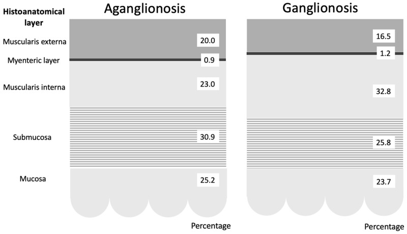
Background/objectives: Intraoperative frozen biopsies are essential during surgery for Hirschsprung's disease (HD). However, this method has several limitations with the need for a faster and real-time diagnostic alternative. For this, consistent histoanatomical and morphometric differences between aganglionic and ganglionic bowel must be established. The primary objective was to compare dimensions of bowel wall layers between aganglionic and ganglionic segments histopathologically in resected rectosigmoid specimens from children with HD. Secondary objectives were to design a diagnostic algorithm to distinguish aganglionosis from ganglionosis and assess whether full bowel wall thickness correlates with patient weight and age.
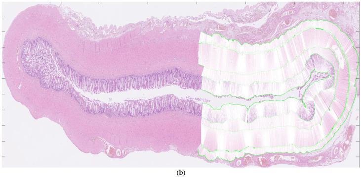
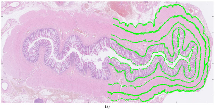
Methods: Each histoanatomic bowel wall layer-mucosa, submucosa, and muscularis propria's layers-was delineated manually on histopathological images. Mean thicknesses were calculated automatically using an in-house image analysis software. Paired parametric tests compared measurements in aganglionic and ganglionic segments.
Results: Resected specimens from 30 children with HD were included. Compared to aganglionic bowel, ganglionic bowel showed a thicker muscularis interna (mean 0.666 mm versus 0.461 mm, CI -0.257-(-0.153), p < 0.001), and a higher muscularis interna/muscularis externa ratio (2.047 mm versus 1.287 mm, CI -0.954-(-0.565), p < 0.001). An algorithm based on these features achieved 100% accuracy in distinguishing aganglionosis from ganglionosis. No significant difference in full bowel wall thickness was found between aganglionic and ganglionic segments, nor any correlation with patient weight or age.
Conclusions: Histoanatomic layer thickness differs between aganglionic and ganglionic bowel, forming the basis of a diagnostic algorithm. Full bowel wall thickness was independent of patient weight and age.

 求助内容:
求助内容: 应助结果提醒方式:


