{"title":"解剖胸主动脉瘤的经典影像学表现及文献回顾。","authors":"S J Ayilara, O Obande, A T Adeniji-Sofoluwe","doi":"","DOIUrl":null,"url":null,"abstract":"<p><strong>Background: </strong>Thoracic aortic aneurysm is an increasingly recognized condition that is sometimes diagnosed incidentally on imaging examinations performed to evaluate other unrelated conditions. Symptomatic presentations of thoracic aortic aneurysms may be due to mass effect on the airway, esophagus, recurrent laryngeal nerve and the thoracic spine. Alternatively, they may present with the dreaded complication of aortic dissection or rupture.Occasionally abnormalities of the aortic contour or size can be detected on routine chest x-ray. However, it is difficult to confidently diagnose thoracic aortic aneurysm on chest x-rays as mediastinal masses may mimic aortic aneurysms. Computed tomography (CT) or magnetic resonance (MR) aortography, with the advantage of obtaining 3D volumetric data, remains the gold standard of imaging with sensitivity and specificity approaching 100%.</p><p><strong>Objective: </strong>To emphasize beauty of cross-sectional images in unraveling confusing opacities on chest radiographs and its sensitivity in identifying potentially lethal chest pathologies.</p><p><strong>Method: </strong>This is a case report of a 58-year-old man with breathlessness and features of congestive cardiac failure. Preliminary chest X-ray revealed a huge soft tissue opacity in the left upper zone of the lung which was conformed as aneurysmal dilatation of the thoracic aorta on chest computed tomography.</p><p><strong>Conclusion: </strong>This case elucidates the importance of cross-sectional imaging in unravelling confusing opacities on chest radiographs and its sensitivity in identifying potentially lethal chest pathologies.</p>","PeriodicalId":72221,"journal":{"name":"Annals of Ibadan postgraduate medicine","volume":"23 1","pages":"121-124"},"PeriodicalIF":0.0000,"publicationDate":"2025-03-31","publicationTypes":"Journal Article","fieldsOfStudy":null,"isOpenAccess":false,"openAccessPdf":"https://www.ncbi.nlm.nih.gov/pmc/articles/PMC12337967/pdf/","citationCount":"0","resultStr":"{\"title\":\"DISSECTING THORACIC AORTIC ANEURYSM WITH CLASSIC IMAGING FINDINGS AND REVIEW OF LITERATURE.\",\"authors\":\"S J Ayilara, O Obande, A T Adeniji-Sofoluwe\",\"doi\":\"\",\"DOIUrl\":null,\"url\":null,\"abstract\":\"<p><strong>Background: </strong>Thoracic aortic aneurysm is an increasingly recognized condition that is sometimes diagnosed incidentally on imaging examinations performed to evaluate other unrelated conditions. Symptomatic presentations of thoracic aortic aneurysms may be due to mass effect on the airway, esophagus, recurrent laryngeal nerve and the thoracic spine. Alternatively, they may present with the dreaded complication of aortic dissection or rupture.Occasionally abnormalities of the aortic contour or size can be detected on routine chest x-ray. However, it is difficult to confidently diagnose thoracic aortic aneurysm on chest x-rays as mediastinal masses may mimic aortic aneurysms. Computed tomography (CT) or magnetic resonance (MR) aortography, with the advantage of obtaining 3D volumetric data, remains the gold standard of imaging with sensitivity and specificity approaching 100%.</p><p><strong>Objective: </strong>To emphasize beauty of cross-sectional images in unraveling confusing opacities on chest radiographs and its sensitivity in identifying potentially lethal chest pathologies.</p><p><strong>Method: </strong>This is a case report of a 58-year-old man with breathlessness and features of congestive cardiac failure. Preliminary chest X-ray revealed a huge soft tissue opacity in the left upper zone of the lung which was conformed as aneurysmal dilatation of the thoracic aorta on chest computed tomography.</p><p><strong>Conclusion: </strong>This case elucidates the importance of cross-sectional imaging in unravelling confusing opacities on chest radiographs and its sensitivity in identifying potentially lethal chest pathologies.</p>\",\"PeriodicalId\":72221,\"journal\":{\"name\":\"Annals of Ibadan postgraduate medicine\",\"volume\":\"23 1\",\"pages\":\"121-124\"},\"PeriodicalIF\":0.0000,\"publicationDate\":\"2025-03-31\",\"publicationTypes\":\"Journal Article\",\"fieldsOfStudy\":null,\"isOpenAccess\":false,\"openAccessPdf\":\"https://www.ncbi.nlm.nih.gov/pmc/articles/PMC12337967/pdf/\",\"citationCount\":\"0\",\"resultStr\":null,\"platform\":\"Semanticscholar\",\"paperid\":null,\"PeriodicalName\":\"Annals of Ibadan postgraduate medicine\",\"FirstCategoryId\":\"1085\",\"ListUrlMain\":\"\",\"RegionNum\":0,\"RegionCategory\":null,\"ArticlePicture\":[],\"TitleCN\":null,\"AbstractTextCN\":null,\"PMCID\":null,\"EPubDate\":\"\",\"PubModel\":\"\",\"JCR\":\"\",\"JCRName\":\"\",\"Score\":null,\"Total\":0}","platform":"Semanticscholar","paperid":null,"PeriodicalName":"Annals of Ibadan postgraduate medicine","FirstCategoryId":"1085","ListUrlMain":"","RegionNum":0,"RegionCategory":null,"ArticlePicture":[],"TitleCN":null,"AbstractTextCN":null,"PMCID":null,"EPubDate":"","PubModel":"","JCR":"","JCRName":"","Score":null,"Total":0}
DISSECTING THORACIC AORTIC ANEURYSM WITH CLASSIC IMAGING FINDINGS AND REVIEW OF LITERATURE.
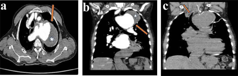
Background: Thoracic aortic aneurysm is an increasingly recognized condition that is sometimes diagnosed incidentally on imaging examinations performed to evaluate other unrelated conditions. Symptomatic presentations of thoracic aortic aneurysms may be due to mass effect on the airway, esophagus, recurrent laryngeal nerve and the thoracic spine. Alternatively, they may present with the dreaded complication of aortic dissection or rupture.Occasionally abnormalities of the aortic contour or size can be detected on routine chest x-ray. However, it is difficult to confidently diagnose thoracic aortic aneurysm on chest x-rays as mediastinal masses may mimic aortic aneurysms. Computed tomography (CT) or magnetic resonance (MR) aortography, with the advantage of obtaining 3D volumetric data, remains the gold standard of imaging with sensitivity and specificity approaching 100%.
Objective: To emphasize beauty of cross-sectional images in unraveling confusing opacities on chest radiographs and its sensitivity in identifying potentially lethal chest pathologies.
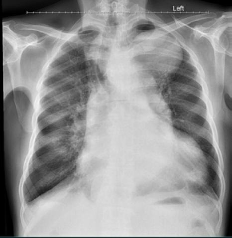
Method: This is a case report of a 58-year-old man with breathlessness and features of congestive cardiac failure. Preliminary chest X-ray revealed a huge soft tissue opacity in the left upper zone of the lung which was conformed as aneurysmal dilatation of the thoracic aorta on chest computed tomography.
Conclusion: This case elucidates the importance of cross-sectional imaging in unravelling confusing opacities on chest radiographs and its sensitivity in identifying potentially lethal chest pathologies.

 求助内容:
求助内容: 应助结果提醒方式:


