Jennifer M McCracken, Gisele A Calderon, Felipe Rivas, Dorothea Erxleben, Taylor Moseley, Lishore A Kumar, Daniel E Kennedy, Swathi Balaji, Adam Hall, Julie C E Hakim
{"title":"揭示阴道纤维化:一种使用博来霉素和上皮破坏的新小鼠模型。","authors":"Jennifer M McCracken, Gisele A Calderon, Felipe Rivas, Dorothea Erxleben, Taylor Moseley, Lishore A Kumar, Daniel E Kennedy, Swathi Balaji, Adam Hall, Julie C E Hakim","doi":"10.4236/ojog.2025.153033","DOIUrl":null,"url":null,"abstract":"<p><p>Vaginal fibrosis induced by cancer therapy continues to take a physical and psychoemotional burden. To advance the efforts to generate a reproducible and cost-effective animal model, we tested the effect of repeated bleomycin instillations with mucosal layer disruption on induction of vaginal fibrosis. Tissue samples collected at various time points were analyzed for fibrosis-related gene expression changes and collagen content. Low (1.5 U/kg) and high-dose (2.5 U/kg) bleomycin instillations alone did not induce fibrosis, but when high-dose bleomycin was combined with epithelial disruption, increased profibrotic gene expression and trichrome staining were observed. To evaluate spatial and temporal changes in the ECM structure and gene expression, tissue samples were collected at 1 day, 3 weeks, and 6 weeks after bleomycin and epithelial disruption. Data analyses revealed a significant decrease in matrix metabolizing genes and an increase in pro-fibrotic genes and inhibitors of matrix metabolizing genes in the bleomycin plus epithelial disruption group at 3 weeks. Elevated levels of the profibrotic genes <i>Acta2</i>, <i>Col1a1</i>, and <i>Col3a</i> were exclusively detected in this group at 3 weeks, and trichrome staining confirmed increased collagen content after 3 weeks. Both average hyaluronan (HA) size and mass distribution were increased 3 weeks after bleomycin plus epithelial disruption with return to baseline by 6 weeks. Epithelial disruption combined with bleomycin induces murine vaginal fibrosis within three weeks, characterized by increased collagen synthesis. The vaginal tissue fully recovers within six weeks, elucidating the regenerative capacity of the vagina.</p>","PeriodicalId":67381,"journal":{"name":"妇产科期刊(英文)","volume":"15 3","pages":"371-386"},"PeriodicalIF":0.0000,"publicationDate":"2025-03-01","publicationTypes":"Journal Article","fieldsOfStudy":null,"isOpenAccess":false,"openAccessPdf":"https://www.ncbi.nlm.nih.gov/pmc/articles/PMC12392699/pdf/","citationCount":"0","resultStr":"{\"title\":\"Unveiling Vaginal Fibrosis: A Novel Murine Model Using Bleomycin and Epithelial Disruption.\",\"authors\":\"Jennifer M McCracken, Gisele A Calderon, Felipe Rivas, Dorothea Erxleben, Taylor Moseley, Lishore A Kumar, Daniel E Kennedy, Swathi Balaji, Adam Hall, Julie C E Hakim\",\"doi\":\"10.4236/ojog.2025.153033\",\"DOIUrl\":null,\"url\":null,\"abstract\":\"<p><p>Vaginal fibrosis induced by cancer therapy continues to take a physical and psychoemotional burden. To advance the efforts to generate a reproducible and cost-effective animal model, we tested the effect of repeated bleomycin instillations with mucosal layer disruption on induction of vaginal fibrosis. Tissue samples collected at various time points were analyzed for fibrosis-related gene expression changes and collagen content. Low (1.5 U/kg) and high-dose (2.5 U/kg) bleomycin instillations alone did not induce fibrosis, but when high-dose bleomycin was combined with epithelial disruption, increased profibrotic gene expression and trichrome staining were observed. To evaluate spatial and temporal changes in the ECM structure and gene expression, tissue samples were collected at 1 day, 3 weeks, and 6 weeks after bleomycin and epithelial disruption. Data analyses revealed a significant decrease in matrix metabolizing genes and an increase in pro-fibrotic genes and inhibitors of matrix metabolizing genes in the bleomycin plus epithelial disruption group at 3 weeks. Elevated levels of the profibrotic genes <i>Acta2</i>, <i>Col1a1</i>, and <i>Col3a</i> were exclusively detected in this group at 3 weeks, and trichrome staining confirmed increased collagen content after 3 weeks. Both average hyaluronan (HA) size and mass distribution were increased 3 weeks after bleomycin plus epithelial disruption with return to baseline by 6 weeks. Epithelial disruption combined with bleomycin induces murine vaginal fibrosis within three weeks, characterized by increased collagen synthesis. The vaginal tissue fully recovers within six weeks, elucidating the regenerative capacity of the vagina.</p>\",\"PeriodicalId\":67381,\"journal\":{\"name\":\"妇产科期刊(英文)\",\"volume\":\"15 3\",\"pages\":\"371-386\"},\"PeriodicalIF\":0.0000,\"publicationDate\":\"2025-03-01\",\"publicationTypes\":\"Journal Article\",\"fieldsOfStudy\":null,\"isOpenAccess\":false,\"openAccessPdf\":\"https://www.ncbi.nlm.nih.gov/pmc/articles/PMC12392699/pdf/\",\"citationCount\":\"0\",\"resultStr\":null,\"platform\":\"Semanticscholar\",\"paperid\":null,\"PeriodicalName\":\"妇产科期刊(英文)\",\"FirstCategoryId\":\"3\",\"ListUrlMain\":\"https://doi.org/10.4236/ojog.2025.153033\",\"RegionNum\":0,\"RegionCategory\":null,\"ArticlePicture\":[],\"TitleCN\":null,\"AbstractTextCN\":null,\"PMCID\":null,\"EPubDate\":\"2025/3/19 0:00:00\",\"PubModel\":\"Epub\",\"JCR\":\"\",\"JCRName\":\"\",\"Score\":null,\"Total\":0}","platform":"Semanticscholar","paperid":null,"PeriodicalName":"妇产科期刊(英文)","FirstCategoryId":"3","ListUrlMain":"https://doi.org/10.4236/ojog.2025.153033","RegionNum":0,"RegionCategory":null,"ArticlePicture":[],"TitleCN":null,"AbstractTextCN":null,"PMCID":null,"EPubDate":"2025/3/19 0:00:00","PubModel":"Epub","JCR":"","JCRName":"","Score":null,"Total":0}
Unveiling Vaginal Fibrosis: A Novel Murine Model Using Bleomycin and Epithelial Disruption.
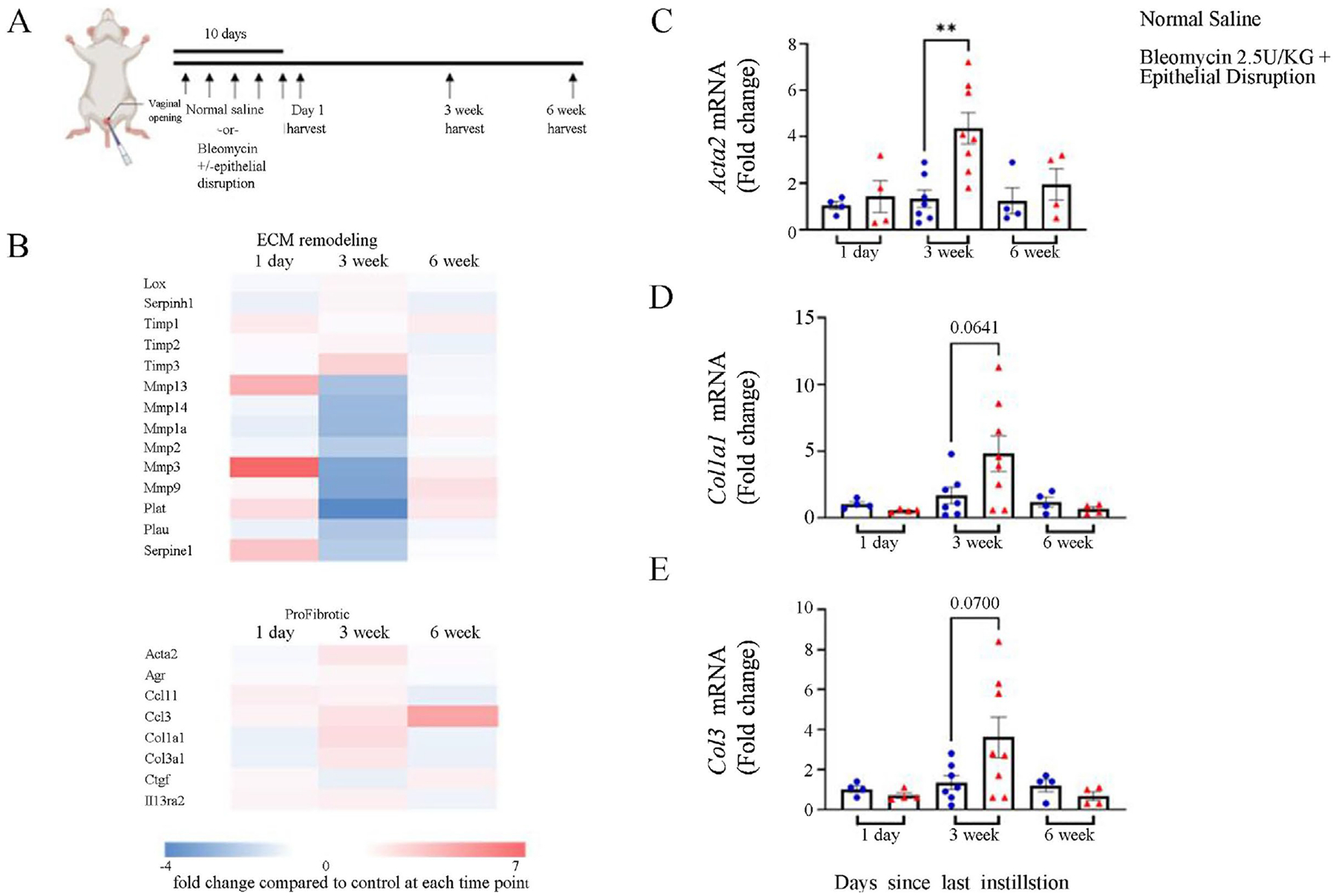
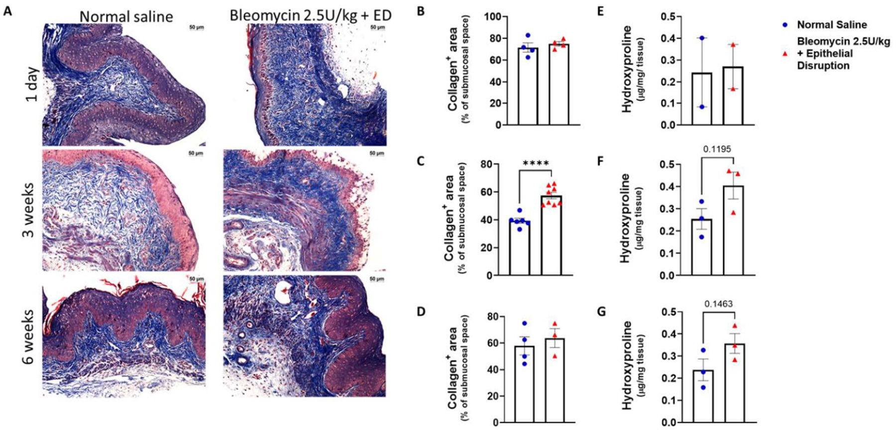
Vaginal fibrosis induced by cancer therapy continues to take a physical and psychoemotional burden. To advance the efforts to generate a reproducible and cost-effective animal model, we tested the effect of repeated bleomycin instillations with mucosal layer disruption on induction of vaginal fibrosis. Tissue samples collected at various time points were analyzed for fibrosis-related gene expression changes and collagen content. Low (1.5 U/kg) and high-dose (2.5 U/kg) bleomycin instillations alone did not induce fibrosis, but when high-dose bleomycin was combined with epithelial disruption, increased profibrotic gene expression and trichrome staining were observed. To evaluate spatial and temporal changes in the ECM structure and gene expression, tissue samples were collected at 1 day, 3 weeks, and 6 weeks after bleomycin and epithelial disruption. Data analyses revealed a significant decrease in matrix metabolizing genes and an increase in pro-fibrotic genes and inhibitors of matrix metabolizing genes in the bleomycin plus epithelial disruption group at 3 weeks. Elevated levels of the profibrotic genes Acta2, Col1a1, and Col3a were exclusively detected in this group at 3 weeks, and trichrome staining confirmed increased collagen content after 3 weeks. Both average hyaluronan (HA) size and mass distribution were increased 3 weeks after bleomycin plus epithelial disruption with return to baseline by 6 weeks. Epithelial disruption combined with bleomycin induces murine vaginal fibrosis within three weeks, characterized by increased collagen synthesis. The vaginal tissue fully recovers within six weeks, elucidating the regenerative capacity of the vagina.
 求助内容:
求助内容: 应助结果提醒方式:


