Weidong Wang, Sheng Chen, Yuan Wang, Cui Jie, Weimin Shen
{"title":"Notch信号对血管瘤干细胞增殖、血管生成和脂肪生成的影响。","authors":"Weidong Wang, Sheng Chen, Yuan Wang, Cui Jie, Weimin Shen","doi":"10.4081/ejh.2025.4241","DOIUrl":null,"url":null,"abstract":"<p><p>Hemangioma-derived stem cells (Hem-SCs) constitute the cellular basis for adipogenesis during infantile hemangioma (IH) regression, with Notch signaling implicated in this process. To elucidate Notch's role in Hem-SCs biology, we isolated primary Hem-SCs from proliferative-phase IH specimens and validated their stem cell characteristics. Three days post-intervention with the γ-secretase inhibitor DAPT (N‑[N‑(3,5‑difluorophenacetyl)‑L‑alanyl]‑S‑phenylglycine t‑butylester), we assessed Notch and PI3K/AKT signaling dynamics while concurrently measuring vascular endothelial growth factor receptor (VEGFR) protein expression. Cellular proliferation was quantified via CCK-8 assay. During adipogenic differentiation (Day 14), RTqPCR evaluated Notch pathway genes (Notch1, Jagged1, Hes1), while adipogenic commitment was determined through Oil Red O staining and adipocyte-specific gene expression (PPARγ, C/EBPα). We demonstrate that DAPT suppresses Notch and PI3K/AKT signaling in Hem-SCs, concomitantly enhancing cellular proliferation and angiogenesis. Simultaneous analysis of VEGFR expression revealed differential DAPT-mediated regulation: VEGFR1 downregulation with concomitant VEGFR2 upregulation. During adipogenic induction, Notch pathway genes (Notch1, Jagged1, Hes1) were significantly downregulated. DAPT treatment further elevated adipogenic markers (PPARγ, C/EBPα) and lipid accumulation. Crucially, co-administration of the PI3K activator 740Y-P reversed DAPT-induced adipogenesis. Mechanistically, Notch inhibition promotes Hem-SCs proliferation, angiogenesis, and adipocyte differentiation by attenuating PI3K/AKT signaling.</p>","PeriodicalId":50487,"journal":{"name":"European Journal of Histochemistry","volume":"69 3","pages":""},"PeriodicalIF":2.1000,"publicationDate":"2025-06-17","publicationTypes":"Journal Article","fieldsOfStudy":null,"isOpenAccess":false,"openAccessPdf":"https://www.ncbi.nlm.nih.gov/pmc/articles/PMC12439523/pdf/","citationCount":"0","resultStr":"{\"title\":\"Effects of Notch signaling on proliferation, angiogenesis, and adipogenesis of hemangioma-derived stem cells.\",\"authors\":\"Weidong Wang, Sheng Chen, Yuan Wang, Cui Jie, Weimin Shen\",\"doi\":\"10.4081/ejh.2025.4241\",\"DOIUrl\":null,\"url\":null,\"abstract\":\"<p><p>Hemangioma-derived stem cells (Hem-SCs) constitute the cellular basis for adipogenesis during infantile hemangioma (IH) regression, with Notch signaling implicated in this process. To elucidate Notch's role in Hem-SCs biology, we isolated primary Hem-SCs from proliferative-phase IH specimens and validated their stem cell characteristics. Three days post-intervention with the γ-secretase inhibitor DAPT (N‑[N‑(3,5‑difluorophenacetyl)‑L‑alanyl]‑S‑phenylglycine t‑butylester), we assessed Notch and PI3K/AKT signaling dynamics while concurrently measuring vascular endothelial growth factor receptor (VEGFR) protein expression. Cellular proliferation was quantified via CCK-8 assay. During adipogenic differentiation (Day 14), RTqPCR evaluated Notch pathway genes (Notch1, Jagged1, Hes1), while adipogenic commitment was determined through Oil Red O staining and adipocyte-specific gene expression (PPARγ, C/EBPα). We demonstrate that DAPT suppresses Notch and PI3K/AKT signaling in Hem-SCs, concomitantly enhancing cellular proliferation and angiogenesis. Simultaneous analysis of VEGFR expression revealed differential DAPT-mediated regulation: VEGFR1 downregulation with concomitant VEGFR2 upregulation. During adipogenic induction, Notch pathway genes (Notch1, Jagged1, Hes1) were significantly downregulated. DAPT treatment further elevated adipogenic markers (PPARγ, C/EBPα) and lipid accumulation. Crucially, co-administration of the PI3K activator 740Y-P reversed DAPT-induced adipogenesis. Mechanistically, Notch inhibition promotes Hem-SCs proliferation, angiogenesis, and adipocyte differentiation by attenuating PI3K/AKT signaling.</p>\",\"PeriodicalId\":50487,\"journal\":{\"name\":\"European Journal of Histochemistry\",\"volume\":\"69 3\",\"pages\":\"\"},\"PeriodicalIF\":2.1000,\"publicationDate\":\"2025-06-17\",\"publicationTypes\":\"Journal Article\",\"fieldsOfStudy\":null,\"isOpenAccess\":false,\"openAccessPdf\":\"https://www.ncbi.nlm.nih.gov/pmc/articles/PMC12439523/pdf/\",\"citationCount\":\"0\",\"resultStr\":null,\"platform\":\"Semanticscholar\",\"paperid\":null,\"PeriodicalName\":\"European Journal of Histochemistry\",\"FirstCategoryId\":\"99\",\"ListUrlMain\":\"https://doi.org/10.4081/ejh.2025.4241\",\"RegionNum\":4,\"RegionCategory\":\"生物学\",\"ArticlePicture\":[],\"TitleCN\":null,\"AbstractTextCN\":null,\"PMCID\":null,\"EPubDate\":\"2025/9/1 0:00:00\",\"PubModel\":\"Epub\",\"JCR\":\"Q4\",\"JCRName\":\"CELL BIOLOGY\",\"Score\":null,\"Total\":0}","platform":"Semanticscholar","paperid":null,"PeriodicalName":"European Journal of Histochemistry","FirstCategoryId":"99","ListUrlMain":"https://doi.org/10.4081/ejh.2025.4241","RegionNum":4,"RegionCategory":"生物学","ArticlePicture":[],"TitleCN":null,"AbstractTextCN":null,"PMCID":null,"EPubDate":"2025/9/1 0:00:00","PubModel":"Epub","JCR":"Q4","JCRName":"CELL BIOLOGY","Score":null,"Total":0}
引用次数: 0
摘要
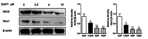
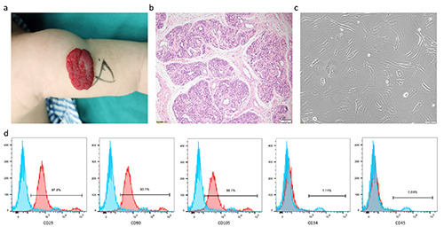
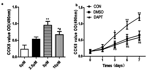
血管瘤来源的干细胞(Hem-SCs)是婴儿血管瘤(IH)消退过程中脂肪形成的细胞基础,Notch信号通路参与了这一过程。为了阐明Notch在Hem-SCs生物学中的作用,我们从增殖期IH标本中分离了原代Hem-SCs,并验证了它们的干细胞特性。在使用γ-分泌酶抑制剂DAPT (N - [N -(3,5 -二氟苯乙酰基)- L -丙烯基]- S -苯甘氨酸-丁酯)干预3天后,我们评估了Notch和PI3K/AKT信号动力学,同时测量了血管内皮生长因子受体(VEGFR)蛋白的表达。CCK-8法定量细胞增殖。在成脂分化期间(第14天),RTqPCR评估Notch通路基因(Notch1, Jagged1, Hes1),而通过油红O染色和脂肪细胞特异性基因表达(PPARγ, C/EBPα)确定成脂承诺。我们发现DAPT抑制Hem-SCs中的Notch和PI3K/AKT信号,同时促进细胞增殖和血管生成。同时对VEGFR表达的分析揭示了dpt介导的差异调控:VEGFR1下调伴随VEGFR2上调。在脂肪诱导过程中,Notch通路基因(Notch1、Jagged1、Hes1)显著下调。DAPT治疗进一步提高了脂肪生成标志物(PPARγ, C/EBPα)和脂质积累。至关重要的是,PI3K激活剂740Y-P的联合使用逆转了dapt诱导的脂肪形成。机制上,Notch抑制通过减弱PI3K/AKT信号传导促进Hem-SCs增殖、血管生成和脂肪细胞分化。
Effects of Notch signaling on proliferation, angiogenesis, and adipogenesis of hemangioma-derived stem cells.
Hemangioma-derived stem cells (Hem-SCs) constitute the cellular basis for adipogenesis during infantile hemangioma (IH) regression, with Notch signaling implicated in this process. To elucidate Notch's role in Hem-SCs biology, we isolated primary Hem-SCs from proliferative-phase IH specimens and validated their stem cell characteristics. Three days post-intervention with the γ-secretase inhibitor DAPT (N‑[N‑(3,5‑difluorophenacetyl)‑L‑alanyl]‑S‑phenylglycine t‑butylester), we assessed Notch and PI3K/AKT signaling dynamics while concurrently measuring vascular endothelial growth factor receptor (VEGFR) protein expression. Cellular proliferation was quantified via CCK-8 assay. During adipogenic differentiation (Day 14), RTqPCR evaluated Notch pathway genes (Notch1, Jagged1, Hes1), while adipogenic commitment was determined through Oil Red O staining and adipocyte-specific gene expression (PPARγ, C/EBPα). We demonstrate that DAPT suppresses Notch and PI3K/AKT signaling in Hem-SCs, concomitantly enhancing cellular proliferation and angiogenesis. Simultaneous analysis of VEGFR expression revealed differential DAPT-mediated regulation: VEGFR1 downregulation with concomitant VEGFR2 upregulation. During adipogenic induction, Notch pathway genes (Notch1, Jagged1, Hes1) were significantly downregulated. DAPT treatment further elevated adipogenic markers (PPARγ, C/EBPα) and lipid accumulation. Crucially, co-administration of the PI3K activator 740Y-P reversed DAPT-induced adipogenesis. Mechanistically, Notch inhibition promotes Hem-SCs proliferation, angiogenesis, and adipocyte differentiation by attenuating PI3K/AKT signaling.
期刊介绍:
The Journal publishes original papers concerning investigations by histochemical and immunohistochemical methods, and performed with the aid of light, super-resolution and electron microscopy, cytometry and imaging techniques. Coverage extends to:
functional cell and tissue biology in animals and plants;
cell differentiation and death;
cell-cell interaction and molecular trafficking;
biology of cell development and senescence;
nerve and muscle cell biology;
cellular basis of diseases.
The histochemical approach is nowadays essentially aimed at locating molecules in the very place where they exert their biological roles, and at describing dynamically specific chemical activities in living cells. Basic research on cell functional organization is essential for understanding the mechanisms underlying major biological processes such as differentiation, the control of tissue homeostasis, and the regulation of normal and tumor cell growth. Even more than in the past, the European Journal of Histochemistry, as a journal of functional cytology, represents the venue where cell scientists may present and discuss their original results, technical improvements and theories.
 求助内容:
求助内容: 应助结果提醒方式:


