Luca Scaravilli, Vincenza Paola Dinuzzi, Felicia Andrei, Andrea Carnevali, Giulia Maria Marini, Martina Pardo, Antonella Nisi, Ildo Scandroglio, Camillo Leonardo Bertoglio
{"title":"三维(3D)血管重建的肠系膜上血管:一个实用的工具,为年轻的外科医生接近右半结肠切除术与CME技术。","authors":"Luca Scaravilli, Vincenza Paola Dinuzzi, Felicia Andrei, Andrea Carnevali, Giulia Maria Marini, Martina Pardo, Antonella Nisi, Ildo Scandroglio, Camillo Leonardo Bertoglio","doi":"10.1186/s12893-025-02945-2","DOIUrl":null,"url":null,"abstract":"<p><strong>Background: </strong>Right hemicolectomy with Complete Mesocolic Excision (CME) and Central Vascular Ligation (CVL) is a complex surgical procedure, partly due to the significant anatomical variability of the superior mesenteric vessels. Three-dimensional (3D) vascular reconstruction, through segmentation-a process that groups pixels with similar characteristics into segments-is a technique that enables the three-dimensional visualization of blood vessels from medical images, usually obtained through CT or magnetic resonance imaging. 3D vascular reconstruction of the superior mesenteric vessels obtained from preoperative images of surgical patients enhances preoperative anatomical understanding, making surgery safer, especially for young surgeons approaching this technique. The primary outcome of this study was to obtain an objective score reflecting surgical residents' understanding of the patient's vascular anatomy from 3D reconstructions of CT images. The secondary outcome was the subjective feedback from senior colorectal surgeons regarding pre-operative use of 3D vascular reconstructions.</p><p><strong>Methods: </strong>A total of 20 patients who underwent right hemicolectomy from 01/10/2023 to 30/09/2024 were included in the study. For each patient, 3D vascular reconstruction was obtained from preoperative CT images. Four surgical residents and two experienced colorectal surgeons were recruited. The residents' understanding of each patient's vascular anatomy was assessed after they looked at the standard pre-operative CT images first and then at their 3D reconstructions respectively. Moreover, the senior colorectal surgeons' opinion on the use of pre-operative 3D vascular reconstruction was assessed.</p><p><strong>Results: </strong>Overall, 3D reconstructions significantly improved residents' anatomical understanding compared to baseline testing (6.71 ± 2.27 vs. 5.26 ± 1.97; p < 0.0001). For three out of four residents examined, 3D vascular reconstruction was statistically superior to standard CT. Colorectal surgeons also gave positive feedback to the use of pre-operative 3D reconstruction.</p><p><strong>Conclusion: </strong>Three-dimensional vascular reconstruction models may help improve surgical trainees' anatomical understanding of mesenteric vascular anatomy compared to conventional CT image interpretation. 3D models may be a useful adjunct to 2D imaging for residents' training and pre-operative planning of CME surgery. More studies are needed to further evaluate the impact of using pre-operative 3D vascular reconstruction.</p>","PeriodicalId":49229,"journal":{"name":"BMC Surgery","volume":"25 1","pages":"378"},"PeriodicalIF":1.8000,"publicationDate":"2025-08-20","publicationTypes":"Journal Article","fieldsOfStudy":null,"isOpenAccess":false,"openAccessPdf":"https://www.ncbi.nlm.nih.gov/pmc/articles/PMC12369134/pdf/","citationCount":"0","resultStr":"{\"title\":\"Three-dimensional (3D) vascular reconstruction of the superior mesenteric vessels: a practical tool for the young surgeon approaching right hemicolectomy with CME technique.\",\"authors\":\"Luca Scaravilli, Vincenza Paola Dinuzzi, Felicia Andrei, Andrea Carnevali, Giulia Maria Marini, Martina Pardo, Antonella Nisi, Ildo Scandroglio, Camillo Leonardo Bertoglio\",\"doi\":\"10.1186/s12893-025-02945-2\",\"DOIUrl\":null,\"url\":null,\"abstract\":\"<p><strong>Background: </strong>Right hemicolectomy with Complete Mesocolic Excision (CME) and Central Vascular Ligation (CVL) is a complex surgical procedure, partly due to the significant anatomical variability of the superior mesenteric vessels. Three-dimensional (3D) vascular reconstruction, through segmentation-a process that groups pixels with similar characteristics into segments-is a technique that enables the three-dimensional visualization of blood vessels from medical images, usually obtained through CT or magnetic resonance imaging. 3D vascular reconstruction of the superior mesenteric vessels obtained from preoperative images of surgical patients enhances preoperative anatomical understanding, making surgery safer, especially for young surgeons approaching this technique. The primary outcome of this study was to obtain an objective score reflecting surgical residents' understanding of the patient's vascular anatomy from 3D reconstructions of CT images. The secondary outcome was the subjective feedback from senior colorectal surgeons regarding pre-operative use of 3D vascular reconstructions.</p><p><strong>Methods: </strong>A total of 20 patients who underwent right hemicolectomy from 01/10/2023 to 30/09/2024 were included in the study. For each patient, 3D vascular reconstruction was obtained from preoperative CT images. Four surgical residents and two experienced colorectal surgeons were recruited. The residents' understanding of each patient's vascular anatomy was assessed after they looked at the standard pre-operative CT images first and then at their 3D reconstructions respectively. Moreover, the senior colorectal surgeons' opinion on the use of pre-operative 3D vascular reconstruction was assessed.</p><p><strong>Results: </strong>Overall, 3D reconstructions significantly improved residents' anatomical understanding compared to baseline testing (6.71 ± 2.27 vs. 5.26 ± 1.97; p < 0.0001). For three out of four residents examined, 3D vascular reconstruction was statistically superior to standard CT. Colorectal surgeons also gave positive feedback to the use of pre-operative 3D reconstruction.</p><p><strong>Conclusion: </strong>Three-dimensional vascular reconstruction models may help improve surgical trainees' anatomical understanding of mesenteric vascular anatomy compared to conventional CT image interpretation. 3D models may be a useful adjunct to 2D imaging for residents' training and pre-operative planning of CME surgery. More studies are needed to further evaluate the impact of using pre-operative 3D vascular reconstruction.</p>\",\"PeriodicalId\":49229,\"journal\":{\"name\":\"BMC Surgery\",\"volume\":\"25 1\",\"pages\":\"378\"},\"PeriodicalIF\":1.8000,\"publicationDate\":\"2025-08-20\",\"publicationTypes\":\"Journal Article\",\"fieldsOfStudy\":null,\"isOpenAccess\":false,\"openAccessPdf\":\"https://www.ncbi.nlm.nih.gov/pmc/articles/PMC12369134/pdf/\",\"citationCount\":\"0\",\"resultStr\":null,\"platform\":\"Semanticscholar\",\"paperid\":null,\"PeriodicalName\":\"BMC Surgery\",\"FirstCategoryId\":\"3\",\"ListUrlMain\":\"https://doi.org/10.1186/s12893-025-02945-2\",\"RegionNum\":3,\"RegionCategory\":\"医学\",\"ArticlePicture\":[],\"TitleCN\":null,\"AbstractTextCN\":null,\"PMCID\":null,\"EPubDate\":\"\",\"PubModel\":\"\",\"JCR\":\"Q2\",\"JCRName\":\"SURGERY\",\"Score\":null,\"Total\":0}","platform":"Semanticscholar","paperid":null,"PeriodicalName":"BMC Surgery","FirstCategoryId":"3","ListUrlMain":"https://doi.org/10.1186/s12893-025-02945-2","RegionNum":3,"RegionCategory":"医学","ArticlePicture":[],"TitleCN":null,"AbstractTextCN":null,"PMCID":null,"EPubDate":"","PubModel":"","JCR":"Q2","JCRName":"SURGERY","Score":null,"Total":0}
Three-dimensional (3D) vascular reconstruction of the superior mesenteric vessels: a practical tool for the young surgeon approaching right hemicolectomy with CME technique.
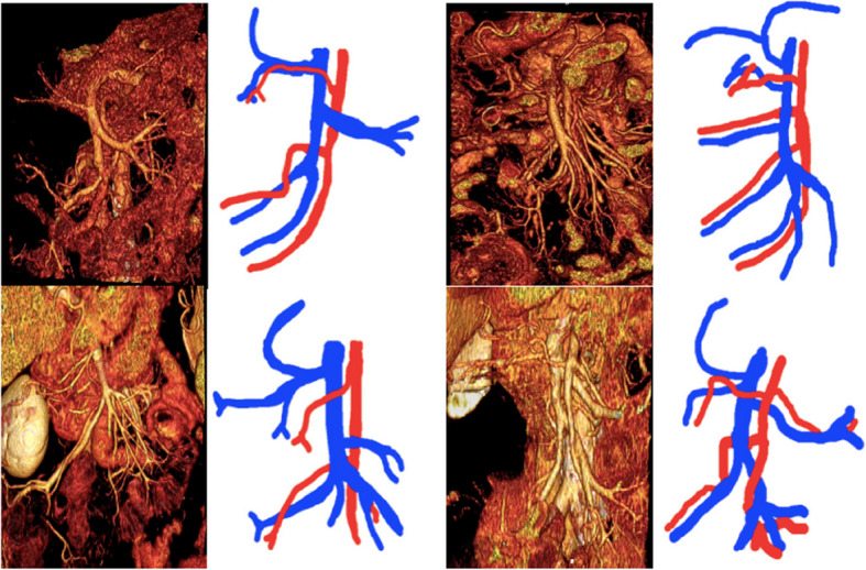
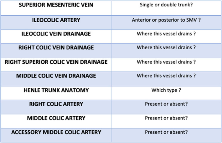
Background: Right hemicolectomy with Complete Mesocolic Excision (CME) and Central Vascular Ligation (CVL) is a complex surgical procedure, partly due to the significant anatomical variability of the superior mesenteric vessels. Three-dimensional (3D) vascular reconstruction, through segmentation-a process that groups pixels with similar characteristics into segments-is a technique that enables the three-dimensional visualization of blood vessels from medical images, usually obtained through CT or magnetic resonance imaging. 3D vascular reconstruction of the superior mesenteric vessels obtained from preoperative images of surgical patients enhances preoperative anatomical understanding, making surgery safer, especially for young surgeons approaching this technique. The primary outcome of this study was to obtain an objective score reflecting surgical residents' understanding of the patient's vascular anatomy from 3D reconstructions of CT images. The secondary outcome was the subjective feedback from senior colorectal surgeons regarding pre-operative use of 3D vascular reconstructions.
Methods: A total of 20 patients who underwent right hemicolectomy from 01/10/2023 to 30/09/2024 were included in the study. For each patient, 3D vascular reconstruction was obtained from preoperative CT images. Four surgical residents and two experienced colorectal surgeons were recruited. The residents' understanding of each patient's vascular anatomy was assessed after they looked at the standard pre-operative CT images first and then at their 3D reconstructions respectively. Moreover, the senior colorectal surgeons' opinion on the use of pre-operative 3D vascular reconstruction was assessed.
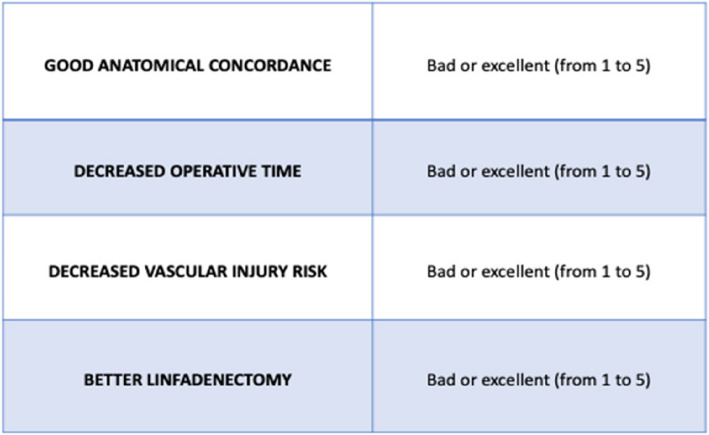
Results: Overall, 3D reconstructions significantly improved residents' anatomical understanding compared to baseline testing (6.71 ± 2.27 vs. 5.26 ± 1.97; p < 0.0001). For three out of four residents examined, 3D vascular reconstruction was statistically superior to standard CT. Colorectal surgeons also gave positive feedback to the use of pre-operative 3D reconstruction.
Conclusion: Three-dimensional vascular reconstruction models may help improve surgical trainees' anatomical understanding of mesenteric vascular anatomy compared to conventional CT image interpretation. 3D models may be a useful adjunct to 2D imaging for residents' training and pre-operative planning of CME surgery. More studies are needed to further evaluate the impact of using pre-operative 3D vascular reconstruction.

 求助内容:
求助内容: 应助结果提醒方式:


