{"title":"胸部超声在肺脓肿诊断及随访中的应用。","authors":"Göksel Menek, Coşkun Doğan","doi":"10.4103/lungindia.lungindia_11_25","DOIUrl":null,"url":null,"abstract":"<p><p>Thoracic ultrasonography (TUS) is a long-established imaging modality with proven efficacy and reliability in the diagnosis of numerous pleuro-parenchymal diseases. Lung abscess (LA) is an infectious disease characterized by liquefactive necrosis and cavitation within the pulmonary parenchyma, typically occurring in immunosuppressed patients or those with various risk factors. Its treatment involves effective antibiotic therapy and, in rare cases, drainage or surgery. Obtaining a sample from the LA, when feasible, is crucial for initiating targeted therapy based on the pathogen identified in the culture. In this case report, we present a 44-year-old female patient hospitalized for an LA. The abscess was visualized via TUS, sampled under ultrasound guidance and successfully managed with ongoing ultrasonographic monitoring.</p>","PeriodicalId":47462,"journal":{"name":"Lung India","volume":"42 5","pages":"461-464"},"PeriodicalIF":1.2000,"publicationDate":"2025-09-01","publicationTypes":"Journal Article","fieldsOfStudy":null,"isOpenAccess":false,"openAccessPdf":"https://www.ncbi.nlm.nih.gov/pmc/articles/PMC12453526/pdf/","citationCount":"0","resultStr":"{\"title\":\"Thoracic ultrasonography in the diagnosis and follow-up of lung abscess.\",\"authors\":\"Göksel Menek, Coşkun Doğan\",\"doi\":\"10.4103/lungindia.lungindia_11_25\",\"DOIUrl\":null,\"url\":null,\"abstract\":\"<p><p>Thoracic ultrasonography (TUS) is a long-established imaging modality with proven efficacy and reliability in the diagnosis of numerous pleuro-parenchymal diseases. Lung abscess (LA) is an infectious disease characterized by liquefactive necrosis and cavitation within the pulmonary parenchyma, typically occurring in immunosuppressed patients or those with various risk factors. Its treatment involves effective antibiotic therapy and, in rare cases, drainage or surgery. Obtaining a sample from the LA, when feasible, is crucial for initiating targeted therapy based on the pathogen identified in the culture. In this case report, we present a 44-year-old female patient hospitalized for an LA. The abscess was visualized via TUS, sampled under ultrasound guidance and successfully managed with ongoing ultrasonographic monitoring.</p>\",\"PeriodicalId\":47462,\"journal\":{\"name\":\"Lung India\",\"volume\":\"42 5\",\"pages\":\"461-464\"},\"PeriodicalIF\":1.2000,\"publicationDate\":\"2025-09-01\",\"publicationTypes\":\"Journal Article\",\"fieldsOfStudy\":null,\"isOpenAccess\":false,\"openAccessPdf\":\"https://www.ncbi.nlm.nih.gov/pmc/articles/PMC12453526/pdf/\",\"citationCount\":\"0\",\"resultStr\":null,\"platform\":\"Semanticscholar\",\"paperid\":null,\"PeriodicalName\":\"Lung India\",\"FirstCategoryId\":\"1085\",\"ListUrlMain\":\"https://doi.org/10.4103/lungindia.lungindia_11_25\",\"RegionNum\":0,\"RegionCategory\":null,\"ArticlePicture\":[],\"TitleCN\":null,\"AbstractTextCN\":null,\"PMCID\":null,\"EPubDate\":\"2025/9/2 0:00:00\",\"PubModel\":\"Epub\",\"JCR\":\"Q4\",\"JCRName\":\"RESPIRATORY SYSTEM\",\"Score\":null,\"Total\":0}","platform":"Semanticscholar","paperid":null,"PeriodicalName":"Lung India","FirstCategoryId":"1085","ListUrlMain":"https://doi.org/10.4103/lungindia.lungindia_11_25","RegionNum":0,"RegionCategory":null,"ArticlePicture":[],"TitleCN":null,"AbstractTextCN":null,"PMCID":null,"EPubDate":"2025/9/2 0:00:00","PubModel":"Epub","JCR":"Q4","JCRName":"RESPIRATORY SYSTEM","Score":null,"Total":0}
Thoracic ultrasonography in the diagnosis and follow-up of lung abscess.
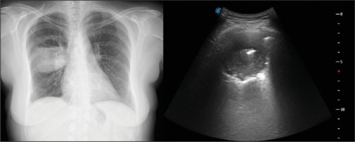
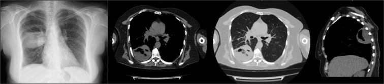
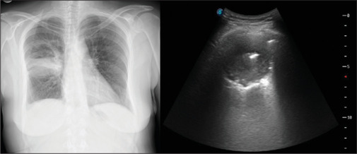
Thoracic ultrasonography (TUS) is a long-established imaging modality with proven efficacy and reliability in the diagnosis of numerous pleuro-parenchymal diseases. Lung abscess (LA) is an infectious disease characterized by liquefactive necrosis and cavitation within the pulmonary parenchyma, typically occurring in immunosuppressed patients or those with various risk factors. Its treatment involves effective antibiotic therapy and, in rare cases, drainage or surgery. Obtaining a sample from the LA, when feasible, is crucial for initiating targeted therapy based on the pathogen identified in the culture. In this case report, we present a 44-year-old female patient hospitalized for an LA. The abscess was visualized via TUS, sampled under ultrasound guidance and successfully managed with ongoing ultrasonographic monitoring.

 求助内容:
求助内容: 应助结果提醒方式:


