{"title":"肺粘膜相关淋巴组织淋巴瘤罕见病例报告。","authors":"Suzhen Yang, Qin Shen","doi":"10.1002/rcr2.70284","DOIUrl":null,"url":null,"abstract":"<p><p>Bronchial mucosa-associated lymphoid tissue is the site of the uncommon malignancy known as mucosa-associated lymphoid tissue (MALT) lymphoma. Due to its lack of distinct clinical signs and imaging characteristics, it is frequently misdiagnosed and underdiagnosed. After receiving unsuccessful treatment for a lung infection or tuberculosis in multiple tertiary care hospitals, we report a male patient who had a solid lesion in his left upper lung for 18 years. The patient was ultimately diagnosed with pulmonary MALT lymphoma at our hospital through a CT-guided percutaneous lung biopsy using the Jiang technique (i.e., employing a laser-assisted guidance system in addition to the conventional CT guidance). It is vital to extend one's perspective, synthesise extrapulmonary signs, and work toward the conditions essential to get a high-quality biopsy specimen when an intrapulmonary lesion is hard to explain by general lung illnesses.</p>","PeriodicalId":45846,"journal":{"name":"Respirology Case Reports","volume":"13 8","pages":"e70284"},"PeriodicalIF":0.8000,"publicationDate":"2025-08-26","publicationTypes":"Journal Article","fieldsOfStudy":null,"isOpenAccess":false,"openAccessPdf":"https://www.ncbi.nlm.nih.gov/pmc/articles/PMC12379836/pdf/","citationCount":"0","resultStr":"{\"title\":\"Rare Case Report of Pulmonary Mucosa-Associated Lymphoid Tissue Lymphoma.\",\"authors\":\"Suzhen Yang, Qin Shen\",\"doi\":\"10.1002/rcr2.70284\",\"DOIUrl\":null,\"url\":null,\"abstract\":\"<p><p>Bronchial mucosa-associated lymphoid tissue is the site of the uncommon malignancy known as mucosa-associated lymphoid tissue (MALT) lymphoma. Due to its lack of distinct clinical signs and imaging characteristics, it is frequently misdiagnosed and underdiagnosed. After receiving unsuccessful treatment for a lung infection or tuberculosis in multiple tertiary care hospitals, we report a male patient who had a solid lesion in his left upper lung for 18 years. The patient was ultimately diagnosed with pulmonary MALT lymphoma at our hospital through a CT-guided percutaneous lung biopsy using the Jiang technique (i.e., employing a laser-assisted guidance system in addition to the conventional CT guidance). It is vital to extend one's perspective, synthesise extrapulmonary signs, and work toward the conditions essential to get a high-quality biopsy specimen when an intrapulmonary lesion is hard to explain by general lung illnesses.</p>\",\"PeriodicalId\":45846,\"journal\":{\"name\":\"Respirology Case Reports\",\"volume\":\"13 8\",\"pages\":\"e70284\"},\"PeriodicalIF\":0.8000,\"publicationDate\":\"2025-08-26\",\"publicationTypes\":\"Journal Article\",\"fieldsOfStudy\":null,\"isOpenAccess\":false,\"openAccessPdf\":\"https://www.ncbi.nlm.nih.gov/pmc/articles/PMC12379836/pdf/\",\"citationCount\":\"0\",\"resultStr\":null,\"platform\":\"Semanticscholar\",\"paperid\":null,\"PeriodicalName\":\"Respirology Case Reports\",\"FirstCategoryId\":\"1085\",\"ListUrlMain\":\"https://doi.org/10.1002/rcr2.70284\",\"RegionNum\":0,\"RegionCategory\":null,\"ArticlePicture\":[],\"TitleCN\":null,\"AbstractTextCN\":null,\"PMCID\":null,\"EPubDate\":\"2025/8/1 0:00:00\",\"PubModel\":\"eCollection\",\"JCR\":\"Q4\",\"JCRName\":\"RESPIRATORY SYSTEM\",\"Score\":null,\"Total\":0}","platform":"Semanticscholar","paperid":null,"PeriodicalName":"Respirology Case Reports","FirstCategoryId":"1085","ListUrlMain":"https://doi.org/10.1002/rcr2.70284","RegionNum":0,"RegionCategory":null,"ArticlePicture":[],"TitleCN":null,"AbstractTextCN":null,"PMCID":null,"EPubDate":"2025/8/1 0:00:00","PubModel":"eCollection","JCR":"Q4","JCRName":"RESPIRATORY SYSTEM","Score":null,"Total":0}
Rare Case Report of Pulmonary Mucosa-Associated Lymphoid Tissue Lymphoma.
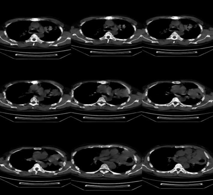
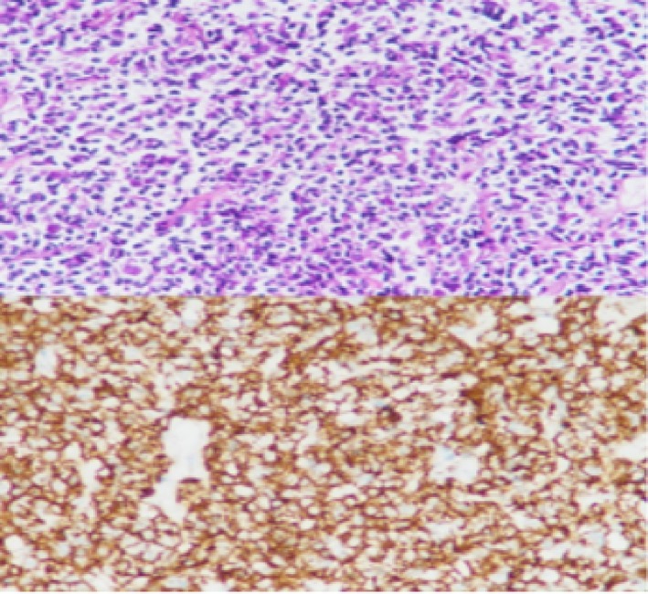
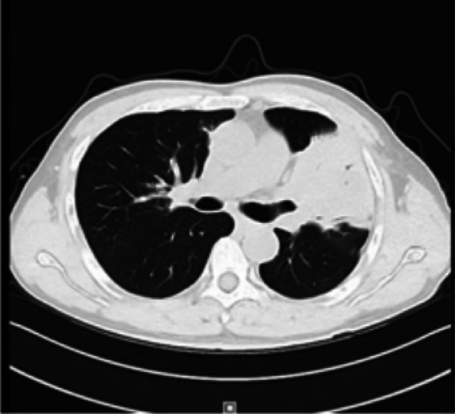
Bronchial mucosa-associated lymphoid tissue is the site of the uncommon malignancy known as mucosa-associated lymphoid tissue (MALT) lymphoma. Due to its lack of distinct clinical signs and imaging characteristics, it is frequently misdiagnosed and underdiagnosed. After receiving unsuccessful treatment for a lung infection or tuberculosis in multiple tertiary care hospitals, we report a male patient who had a solid lesion in his left upper lung for 18 years. The patient was ultimately diagnosed with pulmonary MALT lymphoma at our hospital through a CT-guided percutaneous lung biopsy using the Jiang technique (i.e., employing a laser-assisted guidance system in addition to the conventional CT guidance). It is vital to extend one's perspective, synthesise extrapulmonary signs, and work toward the conditions essential to get a high-quality biopsy specimen when an intrapulmonary lesion is hard to explain by general lung illnesses.
期刊介绍:
Respirology Case Reports is an open-access online journal dedicated to the publication of original clinical case reports, case series, clinical images and clinical videos in all fields of respiratory medicine. The Journal encourages the international exchange between clinicians and researchers of experiences in diagnosing and treating uncommon diseases or diseases with unusual presentations. All manuscripts are peer-reviewed through a streamlined process that aims at providing a rapid turnaround time from submission to publication.

 求助内容:
求助内容: 应助结果提醒方式:


