Siddharth Venkatesh, John S Costanza, Bettye Cox, Chris Finch, Ya Xu
{"title":"尿路上皮癌伴前列腺腺癌同时发生淋巴上皮瘤样和浆细胞样亚型及临床随访。","authors":"Siddharth Venkatesh, John S Costanza, Bettye Cox, Chris Finch, Ya Xu","doi":"10.1155/crip/9068792","DOIUrl":null,"url":null,"abstract":"<p><p>Lymphoepithelioma-like urothelial carcinoma (LELUC) and plasmacytoid urothelial carcinoma (PUC) are rare subtypes. We report a case of simultaneous urothelial carcinoma composed of LELUC and PUC subtypes, along with prostatic adenocarcinoma, with successful clinical management by immunotherapy. The patient, a 54-year-old man with a 40 pack-year smoking history, presented with gross hematuria and dysuria. Imaging revealed focal bladder wall thickening. The patient underwent transurethral resection of bladder tumor (TURBT), followed by cystoprostatectomy. The TURBT revealed LELUC, with muscle invasion. The subsequent cystoprostatectomy specimen displayed a 6.0 cm ulcerative mass, which had focal penetration through the urinary bladder wall. Microscopically, the tumor consisted of sheets of enlarged and pleomorphic tumor cells, mixed with a lymphoplasmacytic infiltrate. Focal plasmacytoid and occasional signet ring cell-like morphologies were observed. Rare tumor cells showed positivity for GATA-3 and p63 immunostains, while the plasmacytoid tumor cells exhibited loss of E-cadherin expression. Additionally, adenocarcinoma of the prostate was present, with a Gleason score of 3 + 3, involving 2% of the prostate tissue. The diagnoses of LELUC, comprising 95% of the tumor, PUC, comprising 5%, and prostatic adenocarcinoma were made. Molecular studies revealed a high tumor mutational burden, and the tumor exhibited PD-L1 expression. The patient received adjuvant immunotherapy with Pembrolizumab and showed no evidence of disease for 3 years up to the time of this report. Morphologic recognition of the various subtypes of urothelial carcinoma, supported by immunohistochemistry, is essential for the proper clinical management of patients. A search of the literature on PubMed revealed no similar cases.</p>","PeriodicalId":45638,"journal":{"name":"Case Reports in Pathology","volume":"2025 ","pages":"9068792"},"PeriodicalIF":0.5000,"publicationDate":"2025-08-21","publicationTypes":"Journal Article","fieldsOfStudy":null,"isOpenAccess":false,"openAccessPdf":"https://www.ncbi.nlm.nih.gov/pmc/articles/PMC12393931/pdf/","citationCount":"0","resultStr":"{\"title\":\"Simultaneous Lymphoepithelioma-Like and Plasmacytoid Subtypes of Urothelial Carcinoma, Along With Prostatic Adenocarcinoma With Clinical Follow-Up.\",\"authors\":\"Siddharth Venkatesh, John S Costanza, Bettye Cox, Chris Finch, Ya Xu\",\"doi\":\"10.1155/crip/9068792\",\"DOIUrl\":null,\"url\":null,\"abstract\":\"<p><p>Lymphoepithelioma-like urothelial carcinoma (LELUC) and plasmacytoid urothelial carcinoma (PUC) are rare subtypes. We report a case of simultaneous urothelial carcinoma composed of LELUC and PUC subtypes, along with prostatic adenocarcinoma, with successful clinical management by immunotherapy. The patient, a 54-year-old man with a 40 pack-year smoking history, presented with gross hematuria and dysuria. Imaging revealed focal bladder wall thickening. The patient underwent transurethral resection of bladder tumor (TURBT), followed by cystoprostatectomy. The TURBT revealed LELUC, with muscle invasion. The subsequent cystoprostatectomy specimen displayed a 6.0 cm ulcerative mass, which had focal penetration through the urinary bladder wall. Microscopically, the tumor consisted of sheets of enlarged and pleomorphic tumor cells, mixed with a lymphoplasmacytic infiltrate. Focal plasmacytoid and occasional signet ring cell-like morphologies were observed. Rare tumor cells showed positivity for GATA-3 and p63 immunostains, while the plasmacytoid tumor cells exhibited loss of E-cadherin expression. Additionally, adenocarcinoma of the prostate was present, with a Gleason score of 3 + 3, involving 2% of the prostate tissue. The diagnoses of LELUC, comprising 95% of the tumor, PUC, comprising 5%, and prostatic adenocarcinoma were made. Molecular studies revealed a high tumor mutational burden, and the tumor exhibited PD-L1 expression. The patient received adjuvant immunotherapy with Pembrolizumab and showed no evidence of disease for 3 years up to the time of this report. Morphologic recognition of the various subtypes of urothelial carcinoma, supported by immunohistochemistry, is essential for the proper clinical management of patients. A search of the literature on PubMed revealed no similar cases.</p>\",\"PeriodicalId\":45638,\"journal\":{\"name\":\"Case Reports in Pathology\",\"volume\":\"2025 \",\"pages\":\"9068792\"},\"PeriodicalIF\":0.5000,\"publicationDate\":\"2025-08-21\",\"publicationTypes\":\"Journal Article\",\"fieldsOfStudy\":null,\"isOpenAccess\":false,\"openAccessPdf\":\"https://www.ncbi.nlm.nih.gov/pmc/articles/PMC12393931/pdf/\",\"citationCount\":\"0\",\"resultStr\":null,\"platform\":\"Semanticscholar\",\"paperid\":null,\"PeriodicalName\":\"Case Reports in Pathology\",\"FirstCategoryId\":\"1085\",\"ListUrlMain\":\"https://doi.org/10.1155/crip/9068792\",\"RegionNum\":0,\"RegionCategory\":null,\"ArticlePicture\":[],\"TitleCN\":null,\"AbstractTextCN\":null,\"PMCID\":null,\"EPubDate\":\"2025/1/1 0:00:00\",\"PubModel\":\"eCollection\",\"JCR\":\"Q4\",\"JCRName\":\"PATHOLOGY\",\"Score\":null,\"Total\":0}","platform":"Semanticscholar","paperid":null,"PeriodicalName":"Case Reports in Pathology","FirstCategoryId":"1085","ListUrlMain":"https://doi.org/10.1155/crip/9068792","RegionNum":0,"RegionCategory":null,"ArticlePicture":[],"TitleCN":null,"AbstractTextCN":null,"PMCID":null,"EPubDate":"2025/1/1 0:00:00","PubModel":"eCollection","JCR":"Q4","JCRName":"PATHOLOGY","Score":null,"Total":0}
Simultaneous Lymphoepithelioma-Like and Plasmacytoid Subtypes of Urothelial Carcinoma, Along With Prostatic Adenocarcinoma With Clinical Follow-Up.
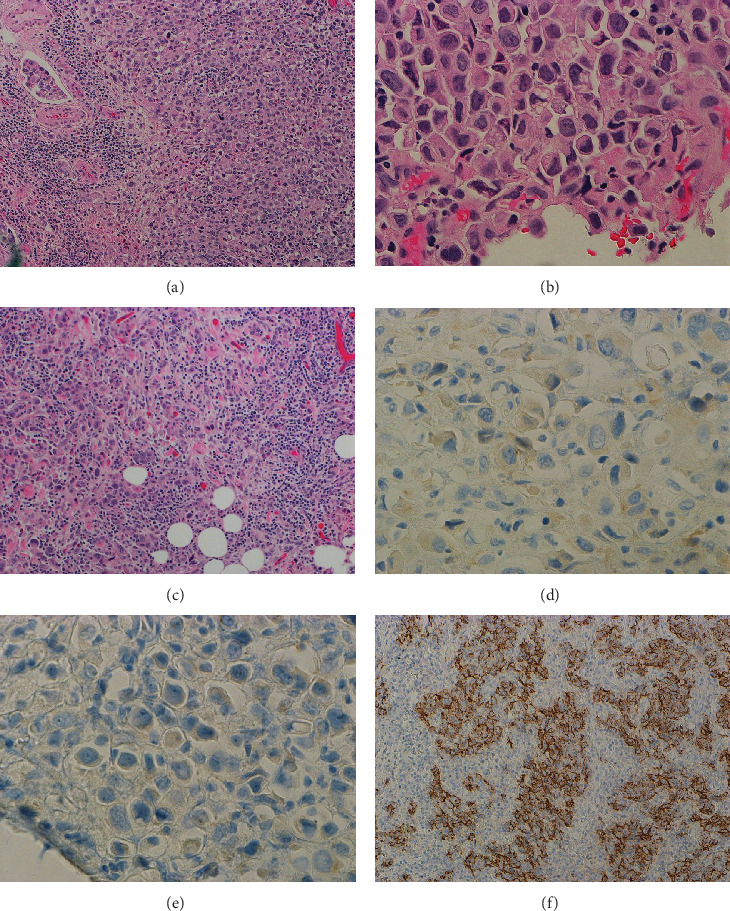
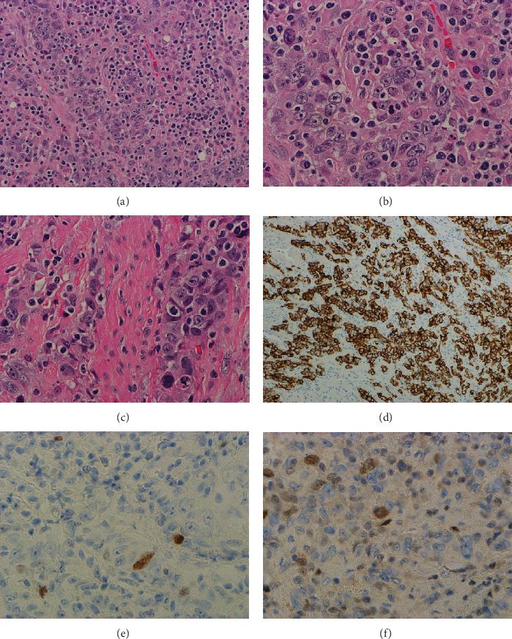
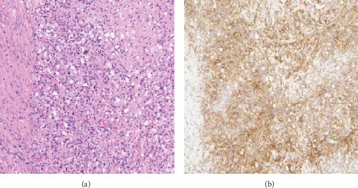
Lymphoepithelioma-like urothelial carcinoma (LELUC) and plasmacytoid urothelial carcinoma (PUC) are rare subtypes. We report a case of simultaneous urothelial carcinoma composed of LELUC and PUC subtypes, along with prostatic adenocarcinoma, with successful clinical management by immunotherapy. The patient, a 54-year-old man with a 40 pack-year smoking history, presented with gross hematuria and dysuria. Imaging revealed focal bladder wall thickening. The patient underwent transurethral resection of bladder tumor (TURBT), followed by cystoprostatectomy. The TURBT revealed LELUC, with muscle invasion. The subsequent cystoprostatectomy specimen displayed a 6.0 cm ulcerative mass, which had focal penetration through the urinary bladder wall. Microscopically, the tumor consisted of sheets of enlarged and pleomorphic tumor cells, mixed with a lymphoplasmacytic infiltrate. Focal plasmacytoid and occasional signet ring cell-like morphologies were observed. Rare tumor cells showed positivity for GATA-3 and p63 immunostains, while the plasmacytoid tumor cells exhibited loss of E-cadherin expression. Additionally, adenocarcinoma of the prostate was present, with a Gleason score of 3 + 3, involving 2% of the prostate tissue. The diagnoses of LELUC, comprising 95% of the tumor, PUC, comprising 5%, and prostatic adenocarcinoma were made. Molecular studies revealed a high tumor mutational burden, and the tumor exhibited PD-L1 expression. The patient received adjuvant immunotherapy with Pembrolizumab and showed no evidence of disease for 3 years up to the time of this report. Morphologic recognition of the various subtypes of urothelial carcinoma, supported by immunohistochemistry, is essential for the proper clinical management of patients. A search of the literature on PubMed revealed no similar cases.

 求助内容:
求助内容: 应助结果提醒方式:


