Chao-Yang Zheng, Jia-Min Zhang, Qian-Sen Lin, Tao Lian, Liang-Pan Shi, Jie-Yun Chen, Ya-Li Cai
{"title":"动态增强磁共振成像放射组学无创预测II/III期直肠癌微卫星不稳定性","authors":"Chao-Yang Zheng, Jia-Min Zhang, Qian-Sen Lin, Tao Lian, Liang-Pan Shi, Jie-Yun Chen, Ya-Li Cai","doi":"10.4251/wjgo.v17.i8.108362","DOIUrl":null,"url":null,"abstract":"<p><strong>Background: </strong>Colorectal cancer stands among the most prevalent digestive system malignancies. The microsatellite instability (MSI) profile plays a crucial role in determining patient outcomes and therapy responsiveness. Traditional MSI evaluation methods require invasive tissue sampling, are lengthy, and can be compromised by intratumoral heterogeneity.</p><p><strong>Aim: </strong>To establish a non-invasive technique utilizing dynamic contrast-enhanced magnetic resonance imaging (DCE-MRI) radiomics and machine learning algorithms to determine MSI status in patients with intermediate-stage rectal cancer.</p><p><strong>Methods: </strong>This retrospective analysis examined 120 individuals diagnosed with stage II/III rectal cancer [30 MSI-high (MSI-H) and 90 microsatellite stability (MSS)/MSI-low (MSI-L) cases]. We extracted comprehensive radiomics signatures from DCE-MRI scans, encompassing textural parameters that reflect tumor heterogeneity, shape-based metrics, and histogram-derived statistical values. Least absolute shrinkage and selection operator regression facilitated feature selection, while predictive frameworks were developed using various classification algorithms (logistic regression, support vector machine, and random forest). Performance assessment utilized separate training and validation cohorts.</p><p><strong>Results: </strong>Our investigation uncovered distinctive imaging characteristics between MSI-H and MSS/MSI-L neoplasms. MSI-H tumors exhibited significantly elevated entropy values (7.84 ± 0.92 <i>vs</i> 6.39 ± 0.83, <i>P</i> = 0.004), enhanced surface-to-volume proportions (0.72 ± 0.14 <i>vs</i> 0.58 ± 0.11, <i>P</i> = 0.008), and heightened signal intensity variation (3642 ± 782 <i>vs</i> 2815 ± 645, <i>P</i> = 0.007). The random forest model demonstrated superior classification capability with area under the curves (AUCs) of 0.891 and 0.896 across training and validation datasets, respectively. An integrated approach combining radiomics with clinical parameters further enhanced performance metrics (AUC 0.923 and 0.914), achieving 88.5% sensitivity alongside 87.2% specificity.</p><p><strong>Conclusion: </strong>DCE-MRI radiomics features interpreted through machine learning frameworks offer an effective strategy for MSI status assessment in intermediate-stage rectal cancer.</p>","PeriodicalId":23762,"journal":{"name":"World Journal of Gastrointestinal Oncology","volume":"17 8","pages":"108362"},"PeriodicalIF":2.5000,"publicationDate":"2025-08-15","publicationTypes":"Journal Article","fieldsOfStudy":null,"isOpenAccess":false,"openAccessPdf":"https://www.ncbi.nlm.nih.gov/pmc/articles/PMC12362526/pdf/","citationCount":"0","resultStr":"{\"title\":\"Noninvasive prediction of microsatellite instability in stage II/III rectal cancer using dynamic contrast-enhanced magnetic resonance imaging radiomics.\",\"authors\":\"Chao-Yang Zheng, Jia-Min Zhang, Qian-Sen Lin, Tao Lian, Liang-Pan Shi, Jie-Yun Chen, Ya-Li Cai\",\"doi\":\"10.4251/wjgo.v17.i8.108362\",\"DOIUrl\":null,\"url\":null,\"abstract\":\"<p><strong>Background: </strong>Colorectal cancer stands among the most prevalent digestive system malignancies. The microsatellite instability (MSI) profile plays a crucial role in determining patient outcomes and therapy responsiveness. Traditional MSI evaluation methods require invasive tissue sampling, are lengthy, and can be compromised by intratumoral heterogeneity.</p><p><strong>Aim: </strong>To establish a non-invasive technique utilizing dynamic contrast-enhanced magnetic resonance imaging (DCE-MRI) radiomics and machine learning algorithms to determine MSI status in patients with intermediate-stage rectal cancer.</p><p><strong>Methods: </strong>This retrospective analysis examined 120 individuals diagnosed with stage II/III rectal cancer [30 MSI-high (MSI-H) and 90 microsatellite stability (MSS)/MSI-low (MSI-L) cases]. We extracted comprehensive radiomics signatures from DCE-MRI scans, encompassing textural parameters that reflect tumor heterogeneity, shape-based metrics, and histogram-derived statistical values. Least absolute shrinkage and selection operator regression facilitated feature selection, while predictive frameworks were developed using various classification algorithms (logistic regression, support vector machine, and random forest). Performance assessment utilized separate training and validation cohorts.</p><p><strong>Results: </strong>Our investigation uncovered distinctive imaging characteristics between MSI-H and MSS/MSI-L neoplasms. MSI-H tumors exhibited significantly elevated entropy values (7.84 ± 0.92 <i>vs</i> 6.39 ± 0.83, <i>P</i> = 0.004), enhanced surface-to-volume proportions (0.72 ± 0.14 <i>vs</i> 0.58 ± 0.11, <i>P</i> = 0.008), and heightened signal intensity variation (3642 ± 782 <i>vs</i> 2815 ± 645, <i>P</i> = 0.007). The random forest model demonstrated superior classification capability with area under the curves (AUCs) of 0.891 and 0.896 across training and validation datasets, respectively. An integrated approach combining radiomics with clinical parameters further enhanced performance metrics (AUC 0.923 and 0.914), achieving 88.5% sensitivity alongside 87.2% specificity.</p><p><strong>Conclusion: </strong>DCE-MRI radiomics features interpreted through machine learning frameworks offer an effective strategy for MSI status assessment in intermediate-stage rectal cancer.</p>\",\"PeriodicalId\":23762,\"journal\":{\"name\":\"World Journal of Gastrointestinal Oncology\",\"volume\":\"17 8\",\"pages\":\"108362\"},\"PeriodicalIF\":2.5000,\"publicationDate\":\"2025-08-15\",\"publicationTypes\":\"Journal Article\",\"fieldsOfStudy\":null,\"isOpenAccess\":false,\"openAccessPdf\":\"https://www.ncbi.nlm.nih.gov/pmc/articles/PMC12362526/pdf/\",\"citationCount\":\"0\",\"resultStr\":null,\"platform\":\"Semanticscholar\",\"paperid\":null,\"PeriodicalName\":\"World Journal of Gastrointestinal Oncology\",\"FirstCategoryId\":\"3\",\"ListUrlMain\":\"https://doi.org/10.4251/wjgo.v17.i8.108362\",\"RegionNum\":4,\"RegionCategory\":\"医学\",\"ArticlePicture\":[],\"TitleCN\":null,\"AbstractTextCN\":null,\"PMCID\":null,\"EPubDate\":\"\",\"PubModel\":\"\",\"JCR\":\"Q2\",\"JCRName\":\"GASTROENTEROLOGY & HEPATOLOGY\",\"Score\":null,\"Total\":0}","platform":"Semanticscholar","paperid":null,"PeriodicalName":"World Journal of Gastrointestinal Oncology","FirstCategoryId":"3","ListUrlMain":"https://doi.org/10.4251/wjgo.v17.i8.108362","RegionNum":4,"RegionCategory":"医学","ArticlePicture":[],"TitleCN":null,"AbstractTextCN":null,"PMCID":null,"EPubDate":"","PubModel":"","JCR":"Q2","JCRName":"GASTROENTEROLOGY & HEPATOLOGY","Score":null,"Total":0}
引用次数: 0
摘要
背景:结直肠癌是最常见的消化系统恶性肿瘤之一。微卫星不稳定性(MSI)在决定患者预后和治疗反应性方面起着至关重要的作用。传统的MSI评估方法需要侵入性组织采样,时间长,并且可能受到肿瘤内异质性的影响。目的:建立一种无创技术,利用动态对比增强磁共振成像(DCE-MRI)放射组学和机器学习算法来确定中期直肠癌患者的MSI状态。方法:回顾性分析120例II/III期直肠癌患者[30例msi高(MSI-H)和90例MSS / msi低(MSI-L)病例]。我们从DCE-MRI扫描中提取了全面的放射组学特征,包括反映肿瘤异质性的纹理参数、基于形状的指标和直方图衍生的统计值。最小绝对收缩和选择算子回归促进了特征选择,而使用各种分类算法(逻辑回归、支持向量机和随机森林)开发了预测框架。绩效评估采用单独的培训和验证队列。结果:我们的研究揭示了MSI-H和MSS/MSI-L肿瘤之间独特的影像学特征。MSI-H肿瘤的熵值显著升高(7.84±0.92 vs 6.39±0.83,P = 0.004),表面体积比显著升高(0.72±0.14 vs 0.58±0.11,P = 0.008),信号强度变化显著升高(3642±782 vs 2815±645,P = 0.007)。随机森林模型在训练集和验证集上的曲线下面积(auc)分别为0.891和0.896,显示出较好的分类能力。放射组学与临床参数相结合的综合方法进一步提高了性能指标(AUC分别为0.923和0.914),灵敏度达到88.5%,特异性为87.2%。结论:通过机器学习框架解释的DCE-MRI放射组学特征为评估中期直肠癌的MSI状态提供了有效的策略。
Noninvasive prediction of microsatellite instability in stage II/III rectal cancer using dynamic contrast-enhanced magnetic resonance imaging radiomics.
Background: Colorectal cancer stands among the most prevalent digestive system malignancies. The microsatellite instability (MSI) profile plays a crucial role in determining patient outcomes and therapy responsiveness. Traditional MSI evaluation methods require invasive tissue sampling, are lengthy, and can be compromised by intratumoral heterogeneity.
Aim: To establish a non-invasive technique utilizing dynamic contrast-enhanced magnetic resonance imaging (DCE-MRI) radiomics and machine learning algorithms to determine MSI status in patients with intermediate-stage rectal cancer.
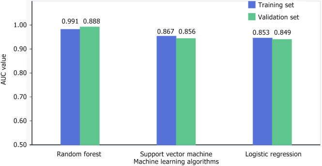
Methods: This retrospective analysis examined 120 individuals diagnosed with stage II/III rectal cancer [30 MSI-high (MSI-H) and 90 microsatellite stability (MSS)/MSI-low (MSI-L) cases]. We extracted comprehensive radiomics signatures from DCE-MRI scans, encompassing textural parameters that reflect tumor heterogeneity, shape-based metrics, and histogram-derived statistical values. Least absolute shrinkage and selection operator regression facilitated feature selection, while predictive frameworks were developed using various classification algorithms (logistic regression, support vector machine, and random forest). Performance assessment utilized separate training and validation cohorts.
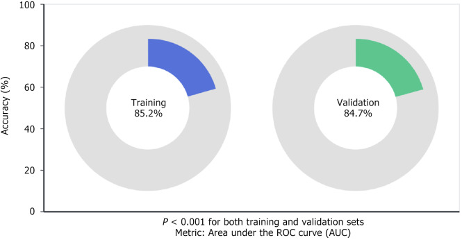
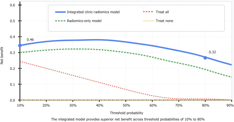
Results: Our investigation uncovered distinctive imaging characteristics between MSI-H and MSS/MSI-L neoplasms. MSI-H tumors exhibited significantly elevated entropy values (7.84 ± 0.92 vs 6.39 ± 0.83, P = 0.004), enhanced surface-to-volume proportions (0.72 ± 0.14 vs 0.58 ± 0.11, P = 0.008), and heightened signal intensity variation (3642 ± 782 vs 2815 ± 645, P = 0.007). The random forest model demonstrated superior classification capability with area under the curves (AUCs) of 0.891 and 0.896 across training and validation datasets, respectively. An integrated approach combining radiomics with clinical parameters further enhanced performance metrics (AUC 0.923 and 0.914), achieving 88.5% sensitivity alongside 87.2% specificity.
Conclusion: DCE-MRI radiomics features interpreted through machine learning frameworks offer an effective strategy for MSI status assessment in intermediate-stage rectal cancer.
期刊介绍:
The World Journal of Gastrointestinal Oncology (WJGO) is a leading academic journal devoted to reporting the latest, cutting-edge research progress and findings of basic research and clinical practice in the field of gastrointestinal oncology.

 求助内容:
求助内容: 应助结果提醒方式:


