Ann Carstens, Geoffrey J Dutton, Hayley J Stannard, Alice Birckhead, William D Barkman, Joanne H Connolly
{"title":"东部灰袋鼠(Macropus giganteus) 16只及红袋鼠(Osphranter rufus) 1只之超声心动图检查。","authors":"Ann Carstens, Geoffrey J Dutton, Hayley J Stannard, Alice Birckhead, William D Barkman, Joanne H Connolly","doi":"10.1111/vru.70079","DOIUrl":null,"url":null,"abstract":"<p><p>Habitat loss, road trauma, predation, disease, and natural disasters impact the health and survival of the family Macropodidae, including kangaroos. Cardiac disease has been reported, including hypertrophic cardiomyopathy (HCM), dilated cardiomyopathy (DCM), nutritional myodegeneration, valvular pathology, cardiovascular parasites, toxoplasmosis, and toxicities. Human research has evaluated macropod pericardium and aortic valves as possible bioprostheses. The goals of this prospective anatomic study were to echocardiographically evaluate opportunistically presented kangaroos: 14 clinically normal eastern grey kangaroos (EGK-Macropus giganteus), two sick EGKs, and one sick red kangaroo (RK, Osphranter rufus). Similar techniques as described in the dog were used. Standard B-mode images, M-mode mensuration, and Doppler measurements were attained; values were descriptively compared with published normal values. The clinically normal animals' M-mode values were similar to the closest weight-related kangaroo values. Most of the animals showed thicker-than-expected left ventricular and interventricular septal walls, and relative wall thickness (RWT) of 0.5 and 0.6; this may be the norm for macropods, but since an RWT>0.45 may indicate human HCM, this should be considered in the kangaroo. The sick animals were euthanized. Necropsy revealed highly suspect HCM in one EGK, and myxomatous mitral valve degeneration with suspect DCM in the other EGK and RK. In conclusion, there are weight-related similarities between previously published kangaroo values. More work is required on a larger number of weight and age cohorts of kangaroos. Subclinical HCM may be present in apparently normal animals. Findings can be used during clinical health assessments and for further research into macropod cardiac conditions.</p>","PeriodicalId":23581,"journal":{"name":"Veterinary Radiology & Ultrasound","volume":"66 5","pages":"e70079"},"PeriodicalIF":1.5000,"publicationDate":"2025-09-01","publicationTypes":"Journal Article","fieldsOfStudy":null,"isOpenAccess":false,"openAccessPdf":"https://www.ncbi.nlm.nih.gov/pmc/articles/PMC12392243/pdf/","citationCount":"0","resultStr":"{\"title\":\"Echocardiographic Examination of 16 Eastern Grey Kangaroos (Macropus giganteus) and One Red Kangaroo (Osphranter rufus).\",\"authors\":\"Ann Carstens, Geoffrey J Dutton, Hayley J Stannard, Alice Birckhead, William D Barkman, Joanne H Connolly\",\"doi\":\"10.1111/vru.70079\",\"DOIUrl\":null,\"url\":null,\"abstract\":\"<p><p>Habitat loss, road trauma, predation, disease, and natural disasters impact the health and survival of the family Macropodidae, including kangaroos. Cardiac disease has been reported, including hypertrophic cardiomyopathy (HCM), dilated cardiomyopathy (DCM), nutritional myodegeneration, valvular pathology, cardiovascular parasites, toxoplasmosis, and toxicities. Human research has evaluated macropod pericardium and aortic valves as possible bioprostheses. The goals of this prospective anatomic study were to echocardiographically evaluate opportunistically presented kangaroos: 14 clinically normal eastern grey kangaroos (EGK-Macropus giganteus), two sick EGKs, and one sick red kangaroo (RK, Osphranter rufus). Similar techniques as described in the dog were used. Standard B-mode images, M-mode mensuration, and Doppler measurements were attained; values were descriptively compared with published normal values. The clinically normal animals' M-mode values were similar to the closest weight-related kangaroo values. Most of the animals showed thicker-than-expected left ventricular and interventricular septal walls, and relative wall thickness (RWT) of 0.5 and 0.6; this may be the norm for macropods, but since an RWT>0.45 may indicate human HCM, this should be considered in the kangaroo. The sick animals were euthanized. Necropsy revealed highly suspect HCM in one EGK, and myxomatous mitral valve degeneration with suspect DCM in the other EGK and RK. In conclusion, there are weight-related similarities between previously published kangaroo values. More work is required on a larger number of weight and age cohorts of kangaroos. Subclinical HCM may be present in apparently normal animals. Findings can be used during clinical health assessments and for further research into macropod cardiac conditions.</p>\",\"PeriodicalId\":23581,\"journal\":{\"name\":\"Veterinary Radiology & Ultrasound\",\"volume\":\"66 5\",\"pages\":\"e70079\"},\"PeriodicalIF\":1.5000,\"publicationDate\":\"2025-09-01\",\"publicationTypes\":\"Journal Article\",\"fieldsOfStudy\":null,\"isOpenAccess\":false,\"openAccessPdf\":\"https://www.ncbi.nlm.nih.gov/pmc/articles/PMC12392243/pdf/\",\"citationCount\":\"0\",\"resultStr\":null,\"platform\":\"Semanticscholar\",\"paperid\":null,\"PeriodicalName\":\"Veterinary Radiology & Ultrasound\",\"FirstCategoryId\":\"97\",\"ListUrlMain\":\"https://doi.org/10.1111/vru.70079\",\"RegionNum\":2,\"RegionCategory\":\"农林科学\",\"ArticlePicture\":[],\"TitleCN\":null,\"AbstractTextCN\":null,\"PMCID\":null,\"EPubDate\":\"\",\"PubModel\":\"\",\"JCR\":\"Q2\",\"JCRName\":\"VETERINARY SCIENCES\",\"Score\":null,\"Total\":0}","platform":"Semanticscholar","paperid":null,"PeriodicalName":"Veterinary Radiology & Ultrasound","FirstCategoryId":"97","ListUrlMain":"https://doi.org/10.1111/vru.70079","RegionNum":2,"RegionCategory":"农林科学","ArticlePicture":[],"TitleCN":null,"AbstractTextCN":null,"PMCID":null,"EPubDate":"","PubModel":"","JCR":"Q2","JCRName":"VETERINARY SCIENCES","Score":null,"Total":0}
Echocardiographic Examination of 16 Eastern Grey Kangaroos (Macropus giganteus) and One Red Kangaroo (Osphranter rufus).
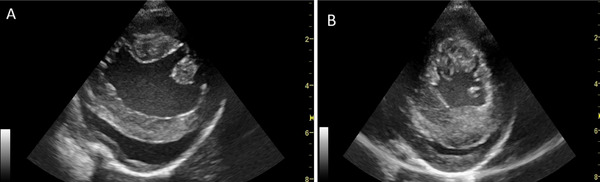
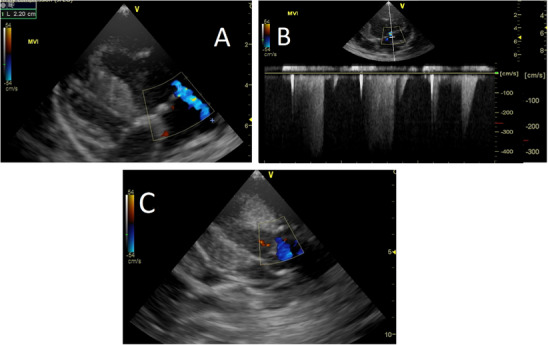
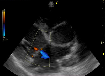
Habitat loss, road trauma, predation, disease, and natural disasters impact the health and survival of the family Macropodidae, including kangaroos. Cardiac disease has been reported, including hypertrophic cardiomyopathy (HCM), dilated cardiomyopathy (DCM), nutritional myodegeneration, valvular pathology, cardiovascular parasites, toxoplasmosis, and toxicities. Human research has evaluated macropod pericardium and aortic valves as possible bioprostheses. The goals of this prospective anatomic study were to echocardiographically evaluate opportunistically presented kangaroos: 14 clinically normal eastern grey kangaroos (EGK-Macropus giganteus), two sick EGKs, and one sick red kangaroo (RK, Osphranter rufus). Similar techniques as described in the dog were used. Standard B-mode images, M-mode mensuration, and Doppler measurements were attained; values were descriptively compared with published normal values. The clinically normal animals' M-mode values were similar to the closest weight-related kangaroo values. Most of the animals showed thicker-than-expected left ventricular and interventricular septal walls, and relative wall thickness (RWT) of 0.5 and 0.6; this may be the norm for macropods, but since an RWT>0.45 may indicate human HCM, this should be considered in the kangaroo. The sick animals were euthanized. Necropsy revealed highly suspect HCM in one EGK, and myxomatous mitral valve degeneration with suspect DCM in the other EGK and RK. In conclusion, there are weight-related similarities between previously published kangaroo values. More work is required on a larger number of weight and age cohorts of kangaroos. Subclinical HCM may be present in apparently normal animals. Findings can be used during clinical health assessments and for further research into macropod cardiac conditions.
期刊介绍:
Veterinary Radiology & Ultrasound is a bimonthly, international, peer-reviewed, research journal devoted to the fields of veterinary diagnostic imaging and radiation oncology. Established in 1958, it is owned by the American College of Veterinary Radiology and is also the official journal for six affiliate veterinary organizations. Veterinary Radiology & Ultrasound is represented on the International Committee of Medical Journal Editors, World Association of Medical Editors, and Committee on Publication Ethics.
The mission of Veterinary Radiology & Ultrasound is to serve as a leading resource for high quality articles that advance scientific knowledge and standards of clinical practice in the areas of veterinary diagnostic radiology, computed tomography, magnetic resonance imaging, ultrasonography, nuclear imaging, radiation oncology, and interventional radiology. Manuscript types include original investigations, imaging diagnosis reports, review articles, editorials and letters to the Editor. Acceptance criteria include originality, significance, quality, reader interest, composition and adherence to author guidelines.

 求助内容:
求助内容: 应助结果提醒方式:


