Jamie Ellis, Mary P Galea, Adam Scheinberg, Peter Simm
{"title":"脊髓疾病儿童外周血定量计算机断层扫描和血液生物标志物。","authors":"Jamie Ellis, Mary P Galea, Adam Scheinberg, Peter Simm","doi":"10.1038/s41394-025-00720-2","DOIUrl":null,"url":null,"abstract":"<p><strong>Study design: </strong>Cross-sectional study OBJECTIVES: Spinal cord disorders (SCD) in children are uncommon, but for those affected the musculoskeletal effects can be severe. Little is known about bone health and pediatric SCD experiences in Australia. We aimed to describe bone and muscle development following pediatric SCD.</p><p><strong>Setting: </strong>The Royal Children's Hospital, Melbourne, Australia METHODS: Ten participants with SCD were recruited and underwent peripheral quantitative computed tomography (pQCT) scans and blood tests to observe bone health biochemistry.</p><p><strong>Results: </strong>Z-scores (mean ± SD) for trabecular density at the 4% tibial site were lower in non-weightbearing children compared to weightbearing children (-6.5 ± 1.5 vs. -2.4 ± 1.5, Total cohort: -5.0 ± 2.6). At the 66% site, muscle cross-sectional area (-4.7 ± 2.2 vs. -1.1 ± 1.7, Total cohort: -3.1 ± 2.7), strength strain index (-3.4 ± 1.3 vs. -1.0 ± 0.4, Total cohort: -2.5 ± 1.6) and total bone cross-sectional area (-2.4 ± 0.8 vs. 0.4 ± 1.7, Total cohort: -1.2 ± 1.9) were also lower in non-weightbearing children. Radial Z-scores revealed reduced total bone area at the 4% site (-3.5 ± 2.1) and strength strain index at the 65% site (-1.3 ± 1.8) in all participants. Serum testing revealed alkaline phosphatase was reduced in three participants, one of whom was also deficient in phosphate and 25-Hydroxyvitamin D.</p><p><strong>Conclusions: </strong>Weightbearing status influenced multiple outcomes including trabecular density, muscle cross-sectional area and bone strength in the tibia.</p>","PeriodicalId":22079,"journal":{"name":"Spinal Cord Series and Cases","volume":"11 1","pages":"24"},"PeriodicalIF":0.9000,"publicationDate":"2025-08-20","publicationTypes":"Journal Article","fieldsOfStudy":null,"isOpenAccess":false,"openAccessPdf":"https://www.ncbi.nlm.nih.gov/pmc/articles/PMC12368188/pdf/","citationCount":"0","resultStr":"{\"title\":\"Peripheral quantitative computed tomography and blood biomarkers in children with spinal cord disorders.\",\"authors\":\"Jamie Ellis, Mary P Galea, Adam Scheinberg, Peter Simm\",\"doi\":\"10.1038/s41394-025-00720-2\",\"DOIUrl\":null,\"url\":null,\"abstract\":\"<p><strong>Study design: </strong>Cross-sectional study OBJECTIVES: Spinal cord disorders (SCD) in children are uncommon, but for those affected the musculoskeletal effects can be severe. Little is known about bone health and pediatric SCD experiences in Australia. We aimed to describe bone and muscle development following pediatric SCD.</p><p><strong>Setting: </strong>The Royal Children's Hospital, Melbourne, Australia METHODS: Ten participants with SCD were recruited and underwent peripheral quantitative computed tomography (pQCT) scans and blood tests to observe bone health biochemistry.</p><p><strong>Results: </strong>Z-scores (mean ± SD) for trabecular density at the 4% tibial site were lower in non-weightbearing children compared to weightbearing children (-6.5 ± 1.5 vs. -2.4 ± 1.5, Total cohort: -5.0 ± 2.6). At the 66% site, muscle cross-sectional area (-4.7 ± 2.2 vs. -1.1 ± 1.7, Total cohort: -3.1 ± 2.7), strength strain index (-3.4 ± 1.3 vs. -1.0 ± 0.4, Total cohort: -2.5 ± 1.6) and total bone cross-sectional area (-2.4 ± 0.8 vs. 0.4 ± 1.7, Total cohort: -1.2 ± 1.9) were also lower in non-weightbearing children. Radial Z-scores revealed reduced total bone area at the 4% site (-3.5 ± 2.1) and strength strain index at the 65% site (-1.3 ± 1.8) in all participants. Serum testing revealed alkaline phosphatase was reduced in three participants, one of whom was also deficient in phosphate and 25-Hydroxyvitamin D.</p><p><strong>Conclusions: </strong>Weightbearing status influenced multiple outcomes including trabecular density, muscle cross-sectional area and bone strength in the tibia.</p>\",\"PeriodicalId\":22079,\"journal\":{\"name\":\"Spinal Cord Series and Cases\",\"volume\":\"11 1\",\"pages\":\"24\"},\"PeriodicalIF\":0.9000,\"publicationDate\":\"2025-08-20\",\"publicationTypes\":\"Journal Article\",\"fieldsOfStudy\":null,\"isOpenAccess\":false,\"openAccessPdf\":\"https://www.ncbi.nlm.nih.gov/pmc/articles/PMC12368188/pdf/\",\"citationCount\":\"0\",\"resultStr\":null,\"platform\":\"Semanticscholar\",\"paperid\":null,\"PeriodicalName\":\"Spinal Cord Series and Cases\",\"FirstCategoryId\":\"1085\",\"ListUrlMain\":\"https://doi.org/10.1038/s41394-025-00720-2\",\"RegionNum\":0,\"RegionCategory\":null,\"ArticlePicture\":[],\"TitleCN\":null,\"AbstractTextCN\":null,\"PMCID\":null,\"EPubDate\":\"\",\"PubModel\":\"\",\"JCR\":\"Q4\",\"JCRName\":\"CLINICAL NEUROLOGY\",\"Score\":null,\"Total\":0}","platform":"Semanticscholar","paperid":null,"PeriodicalName":"Spinal Cord Series and Cases","FirstCategoryId":"1085","ListUrlMain":"https://doi.org/10.1038/s41394-025-00720-2","RegionNum":0,"RegionCategory":null,"ArticlePicture":[],"TitleCN":null,"AbstractTextCN":null,"PMCID":null,"EPubDate":"","PubModel":"","JCR":"Q4","JCRName":"CLINICAL NEUROLOGY","Score":null,"Total":0}
引用次数: 0
摘要
研究设计:横断面研究目的:脊髓疾病(SCD)在儿童中并不常见,但对肌肉骨骼的影响可能是严重的。在澳大利亚,人们对骨骼健康和儿童SCD的经验知之甚少。我们的目的是描述儿童SCD后骨骼和肌肉的发育。方法:招募10名SCD患者,进行外周血定量计算机断层扫描(pQCT)和血液检查,观察骨骼健康生化。结果:与负重儿童相比,非负重儿童4%胫骨部位小梁密度的z分数(平均值±SD)较低(-6.5±1.5比-2.4±1.5,总队列:-5.0±2.6)。在66%部位,非负重儿童的肌肉截面积(-4.7±2.2 vs -1.1±1.7,总队列:-3.1±2.7)、强度应变指数(-3.4±1.3 vs -1.0±0.4,总队列:-2.5±1.6)和总骨截面积(-2.4±0.8 vs. 0.4±1.7,总队列:-1.2±1.9)也较低。径向z分数显示,所有参与者在4%部位的总骨面积(-3.5±2.1)和65%部位的强度应变指数(-1.3±1.8)减少。血清检测显示,三名参与者的碱性磷酸酶降低,其中一人也缺乏磷酸盐和25-羟基维生素d。结论:体重状况影响多种结果,包括胫骨小梁密度、肌肉横截面积和骨强度。
Peripheral quantitative computed tomography and blood biomarkers in children with spinal cord disorders.
Study design: Cross-sectional study OBJECTIVES: Spinal cord disorders (SCD) in children are uncommon, but for those affected the musculoskeletal effects can be severe. Little is known about bone health and pediatric SCD experiences in Australia. We aimed to describe bone and muscle development following pediatric SCD.
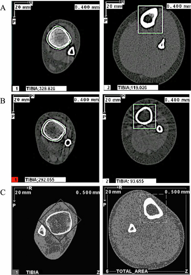
Setting: The Royal Children's Hospital, Melbourne, Australia METHODS: Ten participants with SCD were recruited and underwent peripheral quantitative computed tomography (pQCT) scans and blood tests to observe bone health biochemistry.
Results: Z-scores (mean ± SD) for trabecular density at the 4% tibial site were lower in non-weightbearing children compared to weightbearing children (-6.5 ± 1.5 vs. -2.4 ± 1.5, Total cohort: -5.0 ± 2.6). At the 66% site, muscle cross-sectional area (-4.7 ± 2.2 vs. -1.1 ± 1.7, Total cohort: -3.1 ± 2.7), strength strain index (-3.4 ± 1.3 vs. -1.0 ± 0.4, Total cohort: -2.5 ± 1.6) and total bone cross-sectional area (-2.4 ± 0.8 vs. 0.4 ± 1.7, Total cohort: -1.2 ± 1.9) were also lower in non-weightbearing children. Radial Z-scores revealed reduced total bone area at the 4% site (-3.5 ± 2.1) and strength strain index at the 65% site (-1.3 ± 1.8) in all participants. Serum testing revealed alkaline phosphatase was reduced in three participants, one of whom was also deficient in phosphate and 25-Hydroxyvitamin D.
Conclusions: Weightbearing status influenced multiple outcomes including trabecular density, muscle cross-sectional area and bone strength in the tibia.

 求助内容:
求助内容: 应助结果提醒方式:


