Thejas Marike Shivakumar, Nitika C Panakkal, Shailesh Nayak, Rajagopal Kadavigere, Tanushree R Kamath, Suresh Sukumar
{"title":"BMI正常个体低剂量腹部骨盆计算机断层扫描深度学习重建算法与iDose4图像质量比较","authors":"Thejas Marike Shivakumar, Nitika C Panakkal, Shailesh Nayak, Rajagopal Kadavigere, Tanushree R Kamath, Suresh Sukumar","doi":"10.1177/20503121251336046","DOIUrl":null,"url":null,"abstract":"<p><strong>Objectives: </strong>Radiation exposure has been a cause of concern in computed tomography imaging. Reducing radiation dose increases the image noise which can be compensated by using reconstruction techniques. Recently artificial intelligence-based reconstruction technique has been introduced. Therefore, the purpose of the study was to prospectively compare the image quality between Idose4 and Precise Image in normal BMI individuals.</p><p><strong>Methods: </strong>Sixty-six consecutive patients with a normal body habitus undergoing contrast-enhanced abdomen and pelvis scan were included in the study. All scans were performed using 100 kVp and tube current modulation. The acquired images were reconstructed to iDose4 and precise imaging. Quantitatively images were analyzed by placing regions of interest in different organs to estimate the image noise, signal-to-noise ratio, and contrast-to-noise ratio. Qualitative analysis was done by two radiologists on a five-point Likert scale.</p><p><strong>Results: </strong>Image noise was significantly reduced using Precise Image across the plain (9.11 ± 1.43 vs 8.18 ± 1.2), arterial (14.34 ± 2.1 vs 10.21 ± 1.5), and portovenous phase (14.78 ± 2.30 vs 11.97 ± 2.07) with maximum noise reduction in the arterial and portovenous phases. Signal-to-noise ratio and contrast-to-noise ratio was significantly improved in all the organs across the plain, arterial, and portovenous phases. Qualitative analysis showed no significant difference between Idose4 and Precise Image with regards to visualization of large vessels in the arterial and portovenous phases. However, precise image was graded better than Idose4 with respect to visualization/conspicuity, image noise, and artifacts.</p><p><strong>Conclusion: </strong>Precise Image can be useful in reducing the image noise and improving the signal-to-noise ratio and contrast-to-noise ratio in low-dose computed tomography protocol among normal BMI individuals.</p>","PeriodicalId":21398,"journal":{"name":"SAGE Open Medicine","volume":"13 ","pages":"20503121251336046"},"PeriodicalIF":2.1000,"publicationDate":"2025-08-22","publicationTypes":"Journal Article","fieldsOfStudy":null,"isOpenAccess":false,"openAccessPdf":"https://www.ncbi.nlm.nih.gov/pmc/articles/PMC12374083/pdf/","citationCount":"0","resultStr":"{\"title\":\"A comparison of the image quality between deep learning reconstruction algorithm and iDose4 using low dose abdominopelvic computed tomography for individuals with normal BMI.\",\"authors\":\"Thejas Marike Shivakumar, Nitika C Panakkal, Shailesh Nayak, Rajagopal Kadavigere, Tanushree R Kamath, Suresh Sukumar\",\"doi\":\"10.1177/20503121251336046\",\"DOIUrl\":null,\"url\":null,\"abstract\":\"<p><strong>Objectives: </strong>Radiation exposure has been a cause of concern in computed tomography imaging. Reducing radiation dose increases the image noise which can be compensated by using reconstruction techniques. Recently artificial intelligence-based reconstruction technique has been introduced. Therefore, the purpose of the study was to prospectively compare the image quality between Idose4 and Precise Image in normal BMI individuals.</p><p><strong>Methods: </strong>Sixty-six consecutive patients with a normal body habitus undergoing contrast-enhanced abdomen and pelvis scan were included in the study. All scans were performed using 100 kVp and tube current modulation. The acquired images were reconstructed to iDose4 and precise imaging. Quantitatively images were analyzed by placing regions of interest in different organs to estimate the image noise, signal-to-noise ratio, and contrast-to-noise ratio. Qualitative analysis was done by two radiologists on a five-point Likert scale.</p><p><strong>Results: </strong>Image noise was significantly reduced using Precise Image across the plain (9.11 ± 1.43 vs 8.18 ± 1.2), arterial (14.34 ± 2.1 vs 10.21 ± 1.5), and portovenous phase (14.78 ± 2.30 vs 11.97 ± 2.07) with maximum noise reduction in the arterial and portovenous phases. Signal-to-noise ratio and contrast-to-noise ratio was significantly improved in all the organs across the plain, arterial, and portovenous phases. Qualitative analysis showed no significant difference between Idose4 and Precise Image with regards to visualization of large vessels in the arterial and portovenous phases. However, precise image was graded better than Idose4 with respect to visualization/conspicuity, image noise, and artifacts.</p><p><strong>Conclusion: </strong>Precise Image can be useful in reducing the image noise and improving the signal-to-noise ratio and contrast-to-noise ratio in low-dose computed tomography protocol among normal BMI individuals.</p>\",\"PeriodicalId\":21398,\"journal\":{\"name\":\"SAGE Open Medicine\",\"volume\":\"13 \",\"pages\":\"20503121251336046\"},\"PeriodicalIF\":2.1000,\"publicationDate\":\"2025-08-22\",\"publicationTypes\":\"Journal Article\",\"fieldsOfStudy\":null,\"isOpenAccess\":false,\"openAccessPdf\":\"https://www.ncbi.nlm.nih.gov/pmc/articles/PMC12374083/pdf/\",\"citationCount\":\"0\",\"resultStr\":null,\"platform\":\"Semanticscholar\",\"paperid\":null,\"PeriodicalName\":\"SAGE Open Medicine\",\"FirstCategoryId\":\"1085\",\"ListUrlMain\":\"https://doi.org/10.1177/20503121251336046\",\"RegionNum\":0,\"RegionCategory\":null,\"ArticlePicture\":[],\"TitleCN\":null,\"AbstractTextCN\":null,\"PMCID\":null,\"EPubDate\":\"2025/1/1 0:00:00\",\"PubModel\":\"eCollection\",\"JCR\":\"Q2\",\"JCRName\":\"MEDICINE, GENERAL & INTERNAL\",\"Score\":null,\"Total\":0}","platform":"Semanticscholar","paperid":null,"PeriodicalName":"SAGE Open Medicine","FirstCategoryId":"1085","ListUrlMain":"https://doi.org/10.1177/20503121251336046","RegionNum":0,"RegionCategory":null,"ArticlePicture":[],"TitleCN":null,"AbstractTextCN":null,"PMCID":null,"EPubDate":"2025/1/1 0:00:00","PubModel":"eCollection","JCR":"Q2","JCRName":"MEDICINE, GENERAL & INTERNAL","Score":null,"Total":0}
引用次数: 0
摘要
目的:在计算机断层成像中,辐射暴露一直是一个值得关注的问题。降低辐射剂量会增加图像噪声,而图像噪声可以通过重建技术加以补偿。近年来引入了基于人工智能的重建技术。因此,本研究的目的是前瞻性地比较Idose4和Precise image在BMI正常个体中的图像质量。方法:连续66例身体体质正常的患者行腹部和骨盆增强扫描。所有扫描均使用100 kVp和管电流调制进行。将采集到的图像重构为iDose4并进行精密成像。通过在不同器官上放置感兴趣的区域来定量分析图像,以估计图像的噪声、信噪比和对比噪比。定性分析是由两名放射科医生按照李克特五分制进行的。结果:采用精确成像技术可明显降低平原期(9.11±1.43 vs 8.18±1.2)、动脉期(14.34±2.1 vs 10.21±1.5)和门静脉期(14.78±2.30 vs 11.97±2.07)的图像噪声,其中动脉期和门静脉期降噪最大。平、动脉、门静脉期各脏器的信噪比和信噪比均有明显改善。定性分析显示,Idose4与Precise Image在动脉和门静脉期大血管的显示方面无显著差异。然而,在可视化/显著性、图像噪声和伪影方面,精确图像的评分优于Idose4。结论:在BMI正常人群的低剂量计算机断层扫描中,精确成像有助于降低图像噪声,提高信噪比和对比噪比。
A comparison of the image quality between deep learning reconstruction algorithm and iDose4 using low dose abdominopelvic computed tomography for individuals with normal BMI.
Objectives: Radiation exposure has been a cause of concern in computed tomography imaging. Reducing radiation dose increases the image noise which can be compensated by using reconstruction techniques. Recently artificial intelligence-based reconstruction technique has been introduced. Therefore, the purpose of the study was to prospectively compare the image quality between Idose4 and Precise Image in normal BMI individuals.
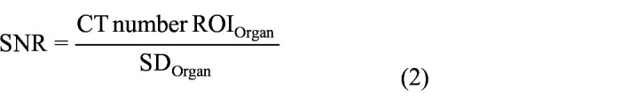
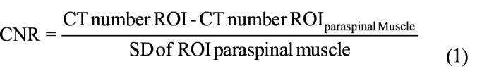
Methods: Sixty-six consecutive patients with a normal body habitus undergoing contrast-enhanced abdomen and pelvis scan were included in the study. All scans were performed using 100 kVp and tube current modulation. The acquired images were reconstructed to iDose4 and precise imaging. Quantitatively images were analyzed by placing regions of interest in different organs to estimate the image noise, signal-to-noise ratio, and contrast-to-noise ratio. Qualitative analysis was done by two radiologists on a five-point Likert scale.
Results: Image noise was significantly reduced using Precise Image across the plain (9.11 ± 1.43 vs 8.18 ± 1.2), arterial (14.34 ± 2.1 vs 10.21 ± 1.5), and portovenous phase (14.78 ± 2.30 vs 11.97 ± 2.07) with maximum noise reduction in the arterial and portovenous phases. Signal-to-noise ratio and contrast-to-noise ratio was significantly improved in all the organs across the plain, arterial, and portovenous phases. Qualitative analysis showed no significant difference between Idose4 and Precise Image with regards to visualization of large vessels in the arterial and portovenous phases. However, precise image was graded better than Idose4 with respect to visualization/conspicuity, image noise, and artifacts.
Conclusion: Precise Image can be useful in reducing the image noise and improving the signal-to-noise ratio and contrast-to-noise ratio in low-dose computed tomography protocol among normal BMI individuals.


 求助内容:
求助内容: 应助结果提醒方式:


